. 2021 May 19;10(5):1254.
doi: 10.3390/cells10051254.

Human Umbilical Cord-Derived Mesenchymal Stem Cells Promote Corneal Epithelial Repair In Vitro

Affiliations

Human Umbilical Cord-Derived Mesenchymal Stem Cells Promote Corneal Epithelial Repair In Vitro

Santhosh Kacham et al. Cells. .

Abstract

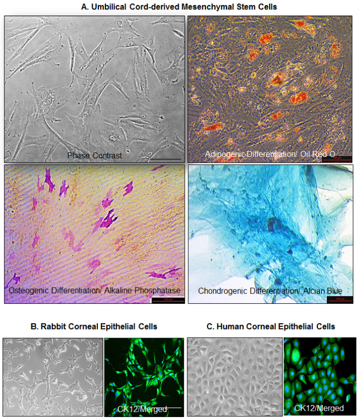
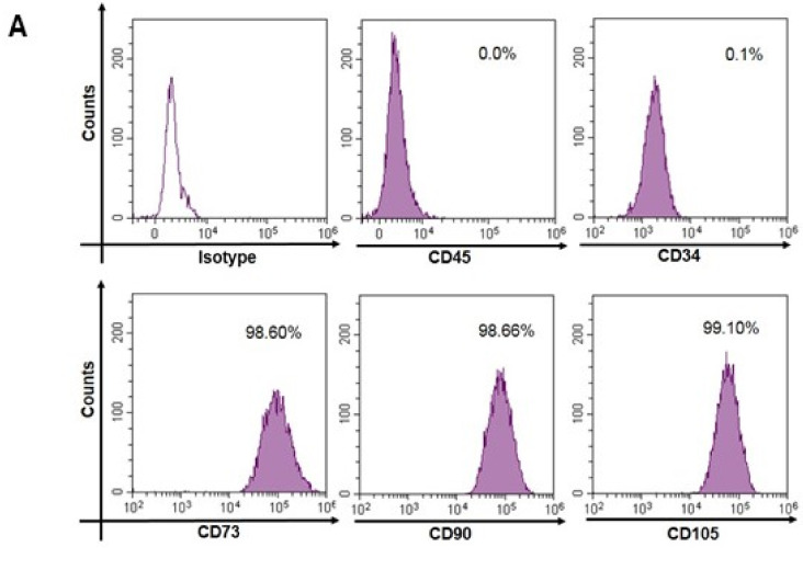
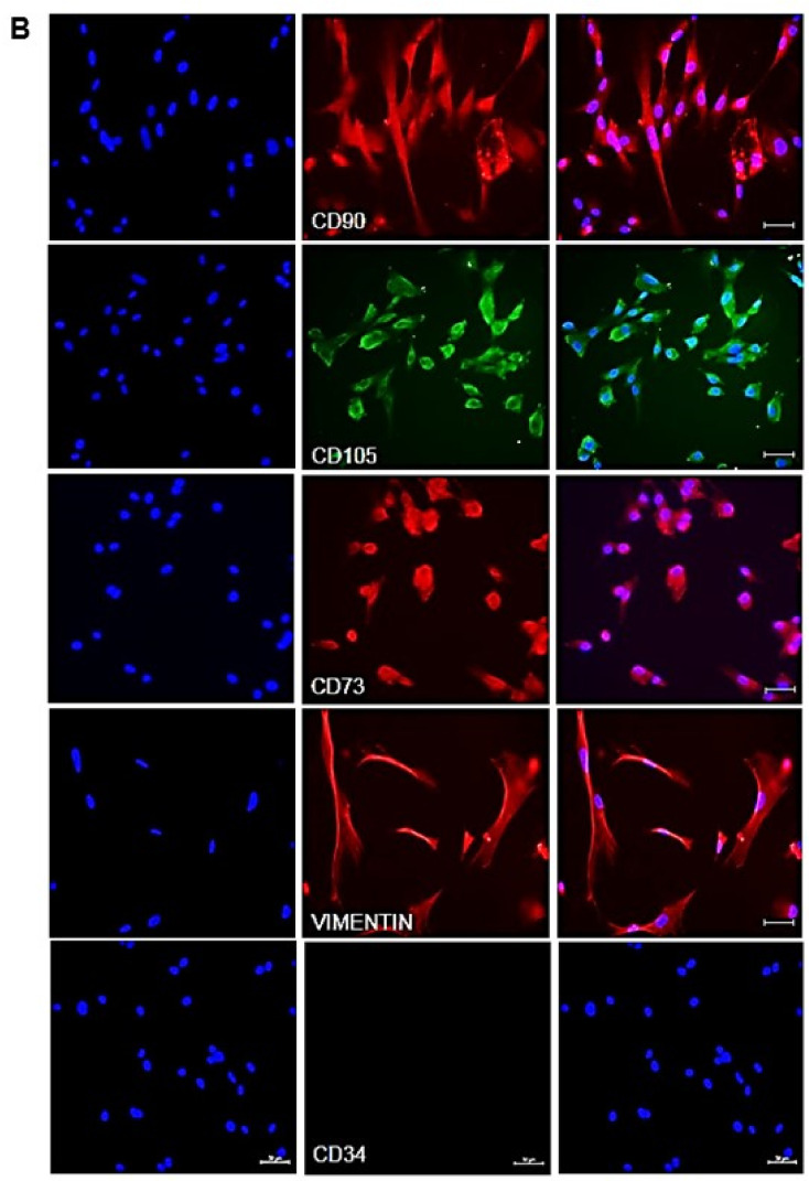
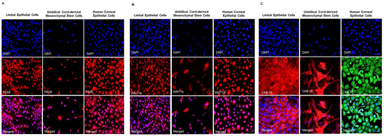
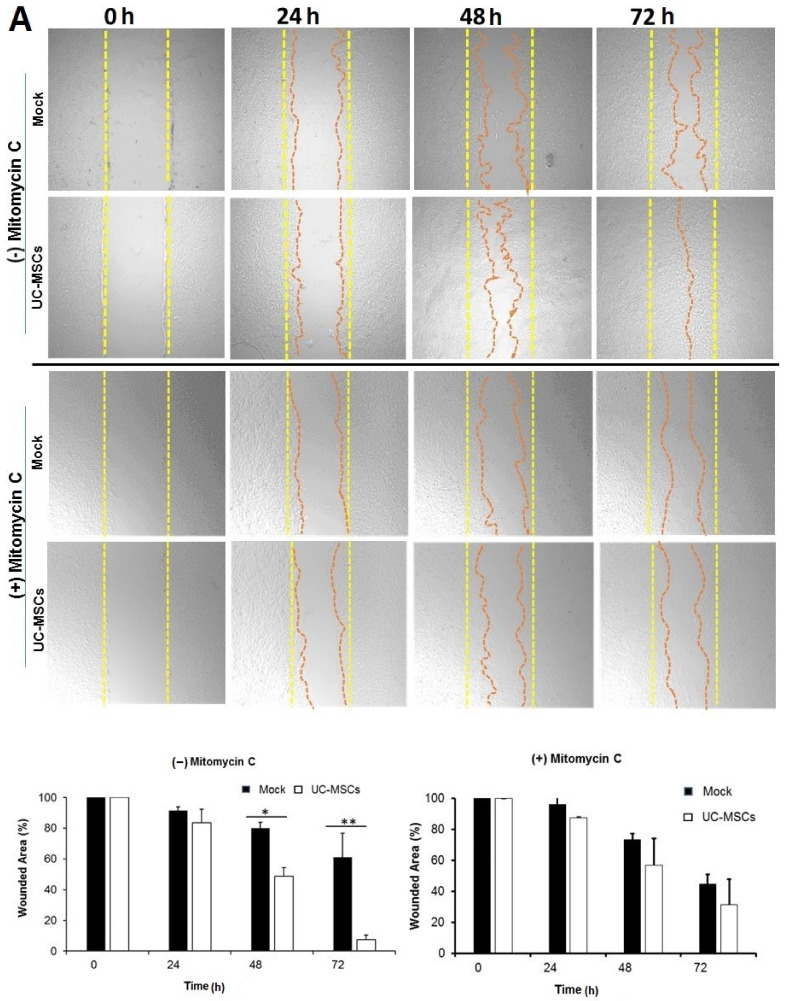
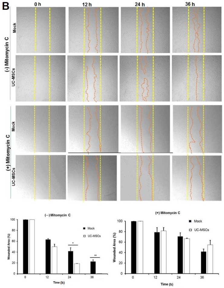
Corneal injuries are among the leading causes of blindness and vision impairment. Trauma, infectious keratitis, thermal and chemical (acids and alkali burn) injuries may lead to irreversible corneal scarring, neovascularization, conjunctivalization, and limbal stem cell deficiency. Bilateral blindness constitutes 12% of total global blindness and corneal transplantation remains a stand-alone treatment modality for the majority of end-stage corneal diseases. However, global shortage of donor corneas, the potential risk of graft rejection, and severe side effects arising from long-term use of immunosuppressive medications, demands alternative therapeutic approaches. Umbilical cord-derived mesenchymal stem cells can be isolated in large numbers using a relatively less invasive procedure. However, their role in injury induced corneal repair is largely unexplored. Here, we isolated, cultured and characterized mesenchymal stem cells from human umbilical cord, and studied the expression of mesenchymal (CD73, CD90, CD105, and CD34), ocular surface and epithelial (PAX6, WNT7A, and CK-8/18) lineage markers through immunofluorescence. The cultured human limbal and corneal epithelial cells were used as controls. Scratch assay was used to study the corneal epithelial repair potential of umbilical cord-derived mesenchymal stem cells, in vitro. The in vitro cultured umbilical cord-derived mesenchymal stem cells were plastic adherent, showed trilineage differentiation and expressed: mesenchymal markers CD90, CD105, CD73; epithelial marker CK-8/18, and ocular lineage developmental markers PAX6 and WNT-7A. Our findings suggest that umbilical cord-derived mesenchymal stem cells promote repair of the injured corneal epithelium by stimulating the proliferation of corneal epithelial cells, in vitro. They may serve as a potential non-ocular source of stem cells for treating injury induced bilateral corneal diseases.

Keywords: cornea; epithelium; limbal stem cell deficiency; mesenchymal stem cells; regeneration; umbilical cord.


Conflict of interest statement

Authors declare that there is no conflict of interest.

Figures

Figure 1
Figure 1
Culture and characterization of cells. (A) Umbilical cord-derived mesenchymal stem cells (UC-MSCs) were cultured in Cytomix medium (Miltenyi Biotec GmbH, Bergisch Gladbach, Germany) and their adipogenic, osteogenic, and chondrogenic differentiation was studied using Oil Red O, Alkaline Phosphatase, and Alcian Blue, respectively. (B) Rabbit corneal epithelial cell line (SIRC) and (C) Human corneal epithelial (HCE) cells were characterized by expression of CK12. Bar size: 100 μm.
Figure 2
Figure 2
Expression of mesenchymal markers by the human umbilical cord-derived mesenchymal stem cells (UC-MSCs). (A) The cultured UC-MSCs showed negative expression of CD45 and CD34 but positive expression of CD73, CD90, and CD105, in flow cytometry. (B) Immunofluorescence was used to study the expression and cellular localization of CD90, CD105, CD73, VIMENTIN, and CD34. The left panel shows DAPI staining, the middle panel shows antigen specific staining and the right panel shows merged image of both DAPI and antigen specific staining. Bar size: 50 μm.
Figure 2
Figure 2
Expression of mesenchymal markers by the human umbilical cord-derived mesenchymal stem cells (UC-MSCs). (A) The cultured UC-MSCs showed negative expression of CD45 and CD34 but positive expression of CD73, CD90, and CD105, in flow cytometry. (B) Immunofluorescence was used to study the expression and cellular localization of CD90, CD105, CD73, VIMENTIN, and CD34. The left panel shows DAPI staining, the middle panel shows antigen specific staining and the right panel shows merged image of both DAPI and antigen specific staining. Bar size: 50 μm.
Figure 3
Figure 3
In vitro cultured human umbilical cord-derived mesenchymal stem cells (UC-MSCs) show endogenous expression of (A) PAX6, an ocular surface developmental marker [40] and a pleiotropic master regulator of eye development [41,42], (B) WNT7A, a key signaling molecule of the Wnt pathway which controls proliferation of human corneal epithelial stem cells [43], and (C) CK-8/18, an epithelial marker [44] under standard culture conditions. This indicates that the hUC-MSCs may possess inherent potential for corneal epithelial repair/regeneration. The human limbal and corneal epithelial cells were used as controls. The top panel shows DAPI staining, the middle panel shows antigen specific staining and the lower panel shows merged image of both DAPI and antigen specific staining in each of the three cell types, respectively. The specificity of the staining was determined by suitable isotype controls (data not shown). Bar size: 50 μm.
Figure 4
Figure 4
Umbilical cord-derived mesenchymal stem cells accelerate corneal epithelial repair by stimulating the proliferation of corneal epithelial cells in vitro. Scratch assay was used as an in vitro model to study the epithelial repair potential of UC-MSCs in cultured (A) rabbit (SIRC) and (B) human (HCE) corneal epithelial cell lines. Mitomycin C treatment (10 μg/mL for 3 h, prior to scratching) was used to inhibit the proliferation of corneal epithelial cells. To mimic epithelial injury, a completely confluent corneal epithelial monolayer was wounded through scratching. Scratched monolayer was incubated with UC-MSCs maintaining indirect contact through culture inserts. Wounded corneal epithelial cells without any incubation with MSCs were used as controls (mock). Epithelial repair was studied through microscopic observations at defined intervals (0, 24, 48, and 72 h in SIRC, and 0, 12, 24, and 36 h in HCE). Total area of the wound/scratch at 0 h was expressed as 100%. Representative data from three independent experiments are shown and values are expressed as mean ± SD (error bar). * p < 0.05, ** p < 0.01.
Figure 4
Figure 4
Umbilical cord-derived mesenchymal stem cells accelerate corneal epithelial repair by stimulating the proliferation of corneal epithelial cells in vitro. Scratch assay was used as an in vitro model to study the epithelial repair potential of UC-MSCs in cultured (A) rabbit (SIRC) and (B) human (HCE) corneal epithelial cell lines. Mitomycin C treatment (10 μg/mL for 3 h, prior to scratching) was used to inhibit the proliferation of corneal epithelial cells. To mimic epithelial injury, a completely confluent corneal epithelial monolayer was wounded through scratching. Scratched monolayer was incubated with UC-MSCs maintaining indirect contact through culture inserts. Wounded corneal epithelial cells without any incubation with MSCs were used as controls (mock). Epithelial repair was studied through microscopic observations at defined intervals (0, 24, 48, and 72 h in SIRC, and 0, 12, 24, and 36 h in HCE). Total area of the wound/scratch at 0 h was expressed as 100%. Representative data from three independent experiments are shown and values are expressed as mean ± SD (error bar). * p < 0.05, ** p < 0.01.

References

    1. Vaidyanathan U., Hopping G.C., Liu H.Y., Somani A.N., Ronquillo Y.C., Hoopes P.C., Moshirfar M. Persistent Corneal Epithelial Defects: A Review Article. Med. Hypothesis Discov. Innov. Ophthalmol. 2019;8:163–176. - PMC - PubMed
    1. DelMonte D.W., Kim T. Anatomy and physiology of the cornea. J. Cataract. Refract. Surg. 2011;37:588–598. doi: 10.1016/j.jcrs.2010.12.037. - DOI - PubMed
    1. Schermer A., Galvin S., Sun T.-T. Differentiation-related expression of a major 64K corneal keratin in vivo and in culture suggests limbal location of corneal epithelial stem cells. J. Cell Biol. 1986;103:49–62. doi: 10.1083/jcb.103.1.49. - DOI - PMC - PubMed
    1. Lavker R.M., Sun T.T. Epithelial stem cells: The eye provides a vision. Eye. 2003;17:937–942. doi: 10.1038/sj.eye.6700575. - DOI - PubMed
    1. Dua H.S., Shanmuganathan A.V., Powell-Richards A., Tighe P.J., Joseph A. Limbal epithelial crypts: A novel anatomical structure and a putative limbal stem cell niche. Br. J. Ophthalmol. 2005;89:529–532. doi: 10.1136/bjo.2004.049742. - DOI - PMC - PubMed

Publication types

MeSH terms