. 2021 May 19;22(10):5363.
doi: 10.3390/ijms22105363.

BPA, BPAF and TMBPF Alter Adipogenesis and Fat Accumulation in Human Mesenchymal Stem Cells, with Implications for Obesity

Affiliations

BPA, BPAF and TMBPF Alter Adipogenesis and Fat Accumulation in Human Mesenchymal Stem Cells, with Implications for Obesity

Isabel C Cohen et al. Int J Mol Sci. .

Abstract

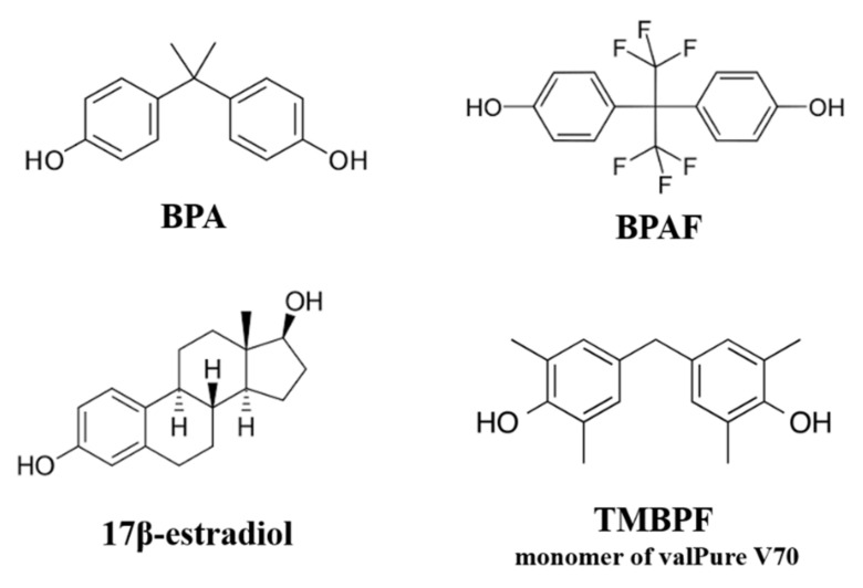
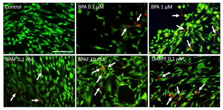
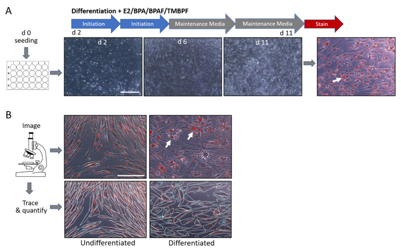
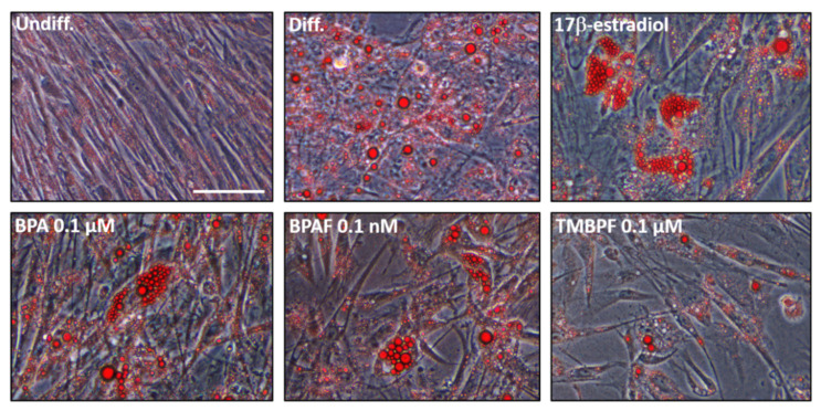
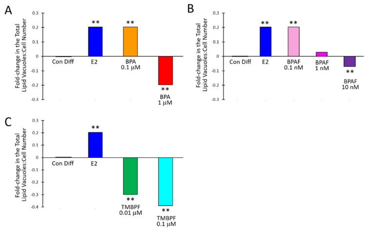
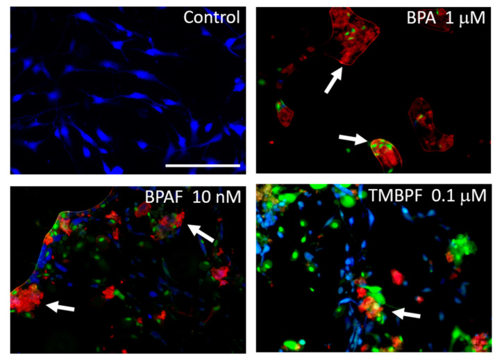
Bisphenol A (BPA) is an endocrine-disrupting chemical used in the production of plastics, and is linked to developmental, reproductive, and metabolic disorders including obesity. Manufacturers have begun using 'BPA-free' alternatives instead of BPA in many consumer products. However, these alternatives have had much less testing and oversight, yet they are already being mass-produced and used across industries from plastics to food-contact coatings. Here, we used human female adipose-derived stem cells (hASCs), a type of adult mesenchymal stem cell, to compare the effects of BPA and BPA alternatives on adipogenesis or fat cell development in vitro. We focused on two commonly used BPA replacements, bisphenol AF (BPAF) and tetramethyl bisphenol F (TMBPF; monomer of the new valPure V70 food-contact coating). Human ASCs were differentiated into adipocytes using chemically defined media in the presence of control differentiation media with and without 17β-estradiol (E2; 10 μM), or with increasing doses of BPA (0, 0.1 and 1 μM), BPAF (0, 0.1, 1 and 10 nM), or TMBPF (0, 0.01 and 0.1 μM). After differentiation, the cells were stained and imaged to visualize and quantify the accumulation of lipid vacuoles and number of developing fat cells. Treated cells were also examined for cell viability and apoptosis (programmed cell death) using the respective cellular assays. Similar to E2, BPA at 0.1 μM and BPAF at 0.1 nM, significantly increased adipogenesis and lipid production by 20% compared to control differentiated cells (based on total lipid vacuole number to cell number ratios), whereas higher levels of BPA and BPAF significantly decreased adipogenesis (p < 0.005). All tested doses of TMBPF significantly reduced adipogenesis and lipid production by 30-40%, likely at least partially through toxic effects on stem cells, as viable cell numbers decreased and apoptosis levels increased throughout differentiation. These findings indicate that low, environmentally-relevant doses of BPA, BPAF, and TMBPF have significant effects on fat cell development and lipid accumulation, with TMBPF having non-estrogenic, anti-adipogenic effects. These and other recent results may provide a potential cellular mechanism between exposure to bisphenols and human obesity, and underscore the likely impact of these chemicals on fat development in vivo.

Keywords: BPA; BPAF; TMBPF; adipogenesis; adipose-derived stem cells (ASCs); endocrine-disrupting chemicals (EDCs); food-contact coating; obesity; plastics; stem cells.


Conflict of interest statement

The authors declare no conflict of interest.

Figures

Figure 1
Figure 1
Chemical structures of BPA, BPA alternatives, and 17β-estradiol.
Figure 2
Figure 2
Cytotoxicity assay of BPA and BPA alternatives. Representative fluorescence images of hASCs treated with BPA at 0.1 µM and 1 µM, BPAF at 0.1 nM and 10 nM, or with TMBPF at 0.1 µM, after 24 h of exposure. Green indicates live cells and red indicates dead cells. Some cell death (see arrows) was found at each dose, especially with 0.1 μM TMBPF, but in general cells remained viable after 24 h of exposure to low-dose BPA and BPAF (200× magnification; scale bar = 150 μM; n = 3–4 slides/treatment; 3 trials).
Figure 3
Figure 3
Timeline and quantification of the differentiation process of human ASCs into adipocytes. (A) Brightfield images of stem cells treated with cycles of initiation media and maintenance media. All cells were fixed, stained with Oil Red-O, imaged, and quantified for lipid vacuoles (white arrows). (B) Representative images of undifferentiated (left) and differentiated (right) cells after staining with Oil Red-O. Tracing of cells and lipid vacuoles in undifferentiated (bottom left) and differentiated (bottom right) cell populations allowed quantification of adipogenesis. Clear morphology changes occur in the differentiated cells compared to undifferentiated controls, as indicated by the round cell shape and lipid vacuoles (arrows in top image and circles in bottom image; 200× magnification; scale bar = 150 μM).
Figure 4
Figure 4
BPA and BPAF increase adipogenesis. Brightfield images of undifferentiated cells, control differentiated cells, and cells treated with either 10 µM 17β-estradiol, 0.1 µM BPA, 0.1 nM BPAF, or 0.1 µM TMBPF during the adipocyte differentiation process. Notice the relatively higher levels of adipogenesis and lipid production in the BPA- and BPAF-differentiated cells, similar to that of 17β-estradiol (see red spheres indicating large lipid vacuoles; 400× magnification; scale bar = 75 μM).
Figure 5
Figure 5
BPA and alternatives alter adipogenesis. The fold-change in the total ratio of lipid vacuoles to cell number in comparison to control differentiated cells (set to 0) after 11 days of differentiation and exposure to increasing doses of (A) BPA, (B) BPAF, or (C) TMBPF (n = 47–103 images/treatment; 4–10 wells/treatment; 2–3 trials; ** p < 0.005).
Figure 6
Figure 6
BPA and alternatives increase apoptosis. Representative fluorescent images of control, BPA-, BPAF-, and TMBPF-treated cells examined with an apoptosis-necrosis assay. Some cells treated with BPA, BPAF, and TMBPF exhibit clear signs of apoptosis and necrosis (see arrows; red = Apopxin Deep Red, indicates apoptosis; green = DNA Nuclear Green DCS1, indicates late-stage apoptosis and necrosis; blue = CytoCalcein Violet 450, indicates normal live cells) (200× magnification; scale bar = 150 μM).

Similar articles

Cited by

References

    1. Vogel S.A. The politics of plastics: The making and unmaking of Bisphenol A “Safety”. Am. J. Public Health. 2009;99:559–566. doi: 10.2105/AJPH.2008.159228. - DOI - PMC - PubMed
    1. Janesick A., Bloomberg B. Obesogens, stem cells, and the developmental programming of obesity. Int. J. Androl. 2012;35:437–448. doi: 10.1111/j.1365-2605.2012.01247.x. - DOI - PMC - PubMed
    1. Braun J.M., Hauser R. Bisphenol A and children’s health. Curr. Opin. Pediatr. 2011;23:233–239. doi: 10.1097/MOP.0b013e3283445675. - DOI - PMC - PubMed
    1. Flint S., Markle T., Thompson S., Wallace E. Bisphenol A exposure, effects, and policy: A wildlife perspective. J. Environ. Manag. 2012;104:19–34. doi: 10.1016/j.jenvman.2012.03.021. - DOI - PubMed
    1. Onundi Y., Drake B.A., Malecky R.T., DeNardo M.A., Mills M.R., Kundu S., Ryabov A.D., Beach E.S., Horwitz C.P., Simonich M.T., et al. A multidisciplinary investigation of the technical and environmental performances of TAML/peroxide elimination of Bisphenol A compounds from water. Green Chem. 2017;19:4234–4262. doi: 10.1039/C7GC01415E. - DOI

LinkOut - more resources