. 2021 May 18;11(5):158.
doi: 10.3390/bios11050158.

A Self-Priming Microfluidic Chip with Cushion Chambers for Easy Digital PCR

Affiliations

A Self-Priming Microfluidic Chip with Cushion Chambers for Easy Digital PCR

Gangwei Xu et al. Biosensors (Basel). .

Abstract

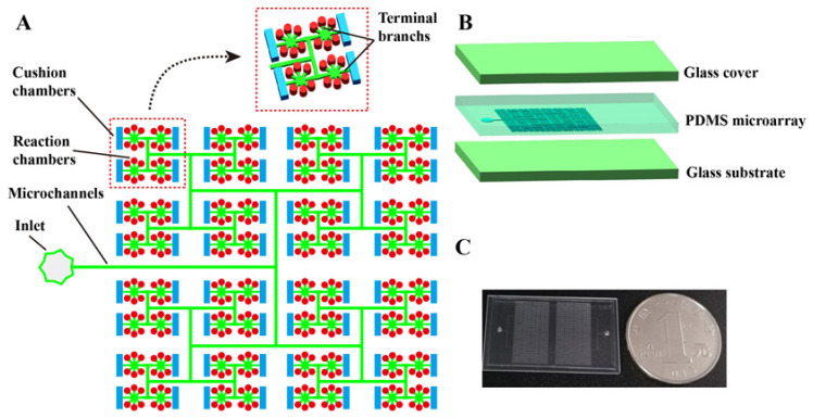
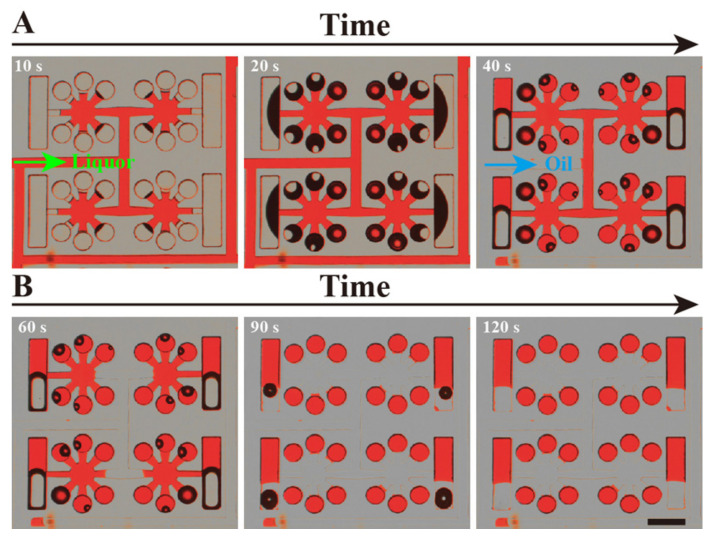
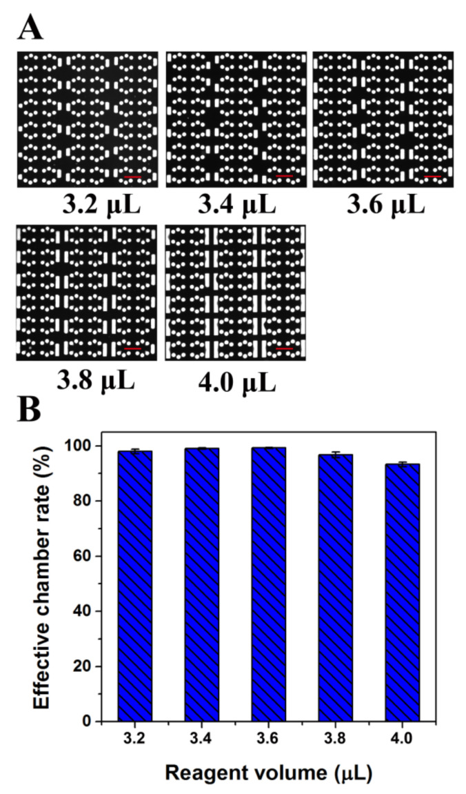
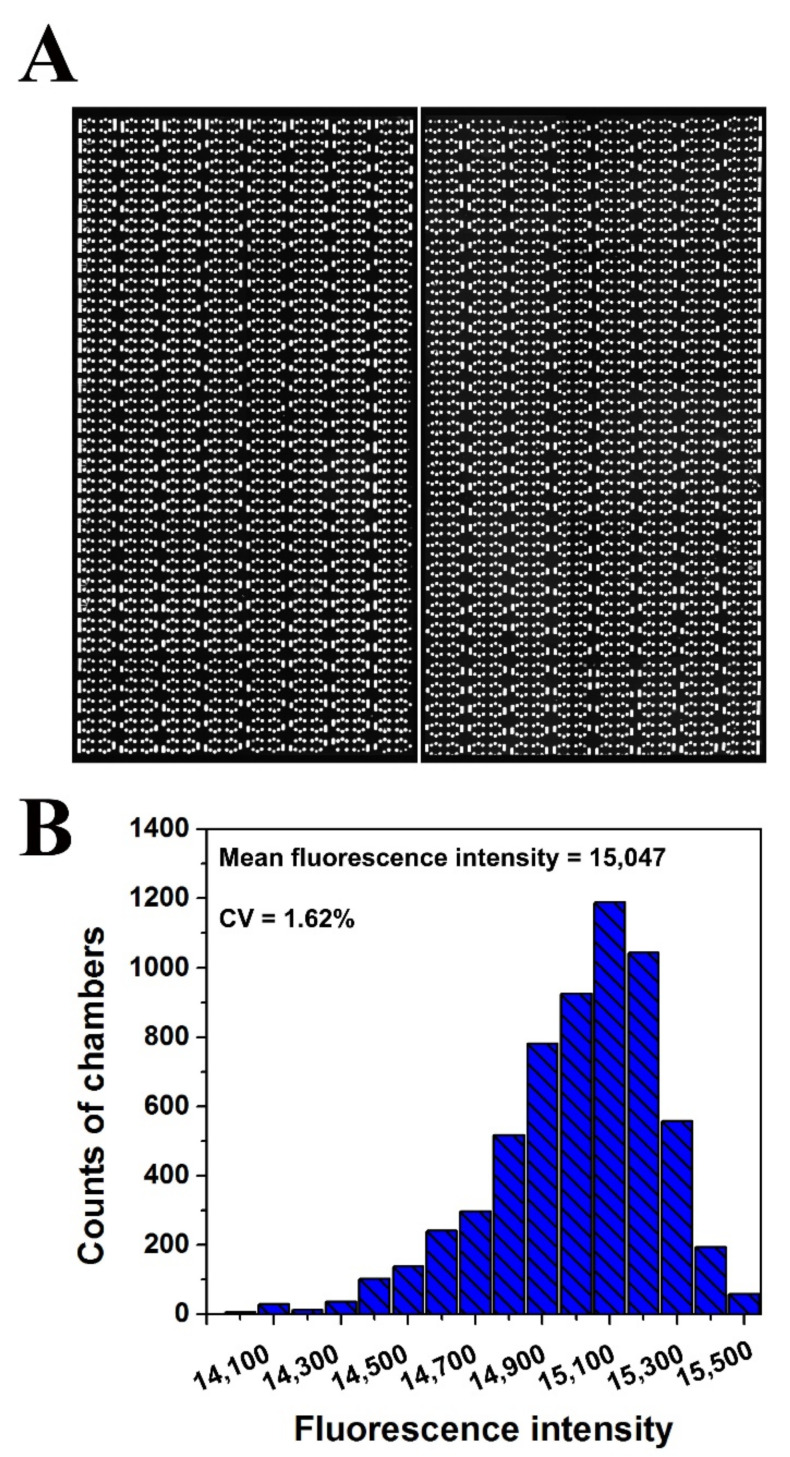
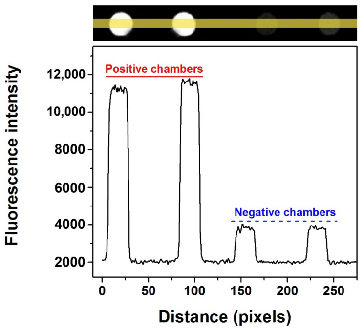
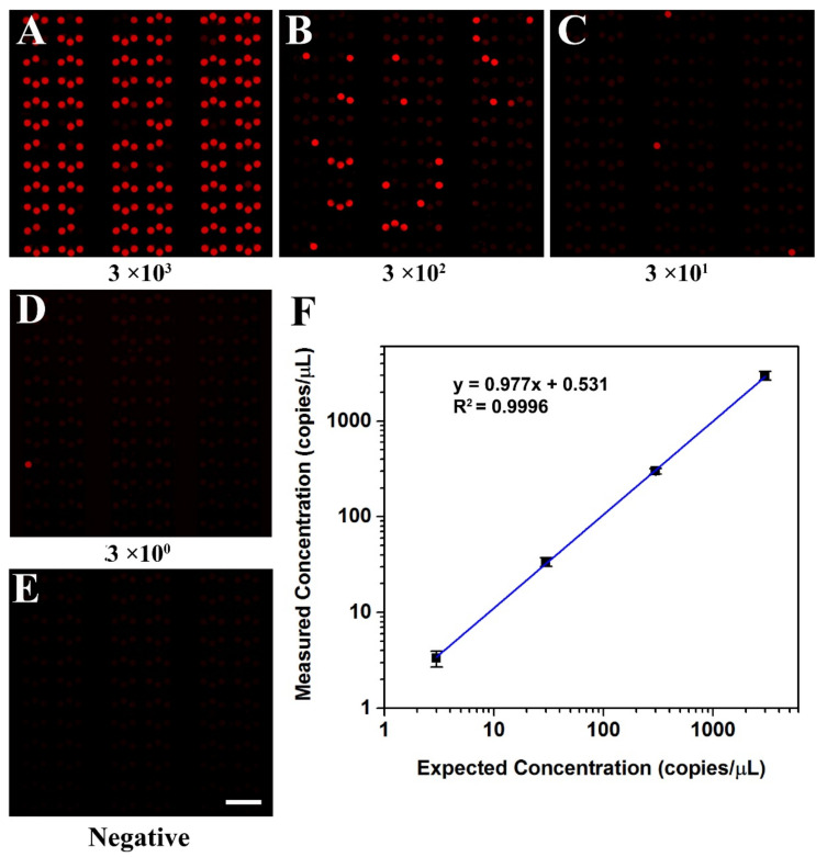
A polydimethylsiloxane (PDMS)-based self-priming microfluidic chip with cushion chambers is presented in this study for robust and easy-operation digital polymerase chain reaction (dPCR). The chip has only one inlet and can partition samples autonomously through negative pressure, provided by a de-gassed PDMS layer with a multi-level vertical branching microchannel design. Meanwhile, cushion chambers make the chip capable of very robust use for sample partitioning. Finally, the proposed microfluidic chip showed excellent performance in the absolute quantification of a target gene by performing quantitative detection of a 10-fold serial dilution DNA template. Owing to its characteristics of easy operation, low cost, and high robustness, the proposed dPCR chip is expected to further promote the extensive application of digital PCR, especially in resource-limited settings.

Keywords: cushion chambers; digital PCR; microfluidic chip; self-priming.


Conflict of interest statement

The authors declare no conflict of interest.

Figures

Figure 1
Figure 1
Schematic of self-priming dPCR chip with cushion chambers. (A) Schematic of the dPCR chip structural design. The chip contains two types of chambers with different volumes and shapes. The chip has only one inlet, without any outlets. The main channel is divided vertically into many branches, step by step. The end of each branch is connected with six reaction chambers and one cushion chamber. (B) Schematic diagram of the layered structure of the dPCR chip, which is composed of two layers of glass coverslips and the PDMS (inlet and microarray-layer) sandwiched between them. (C) Photograph of the prototype dPCR chip. The chip contains two panels, and each panel has 3072 reaction chambers and 512 cushion chambers. The size of the chip is 20 mm × 35 mm.
Figure 2
Figure 2
Optical micrograph of the process of red dye and oil self-primed into the dPCR chip. (A) The red dye solution (liquor) was sucked into the microchannel and chambers. (B) The oil (transparent) phase flowed into the chip and separated the red dye solution. Lastly, the oil phase entirely separates the red dye in each chamber. The black shadow in the chambers is air-water interface.
Figure 3
Figure 3
Diagram of the cushioning effect analysis of the dPCR chip. (A) Fluorescent image of reaction chambers filled with fluorescent solution after loading different amount of fluorescent reagent. (B) Effective chamber rate after loading different amount of fluorescent reagent. (All scale bars represent 400 μm).
Figure 4
Figure 4
Diagram of the uniformity analysis of the dPCR chip. (A) Fluorescent image of reaction chambers filled with fluorescent solution. (B) Histogram of the fluorescence intensities distribution of all the reaction chambers (6144) in one chip.
Figure 5
Figure 5
Fluorescence intensity contrast between positive and negative chambers of KRAS wild probes-CY5 with self-priming after PCR amplification. (The yellow line represents the statistical area of fluorescence intensity. The black curve below shows the change of fluorescence intensity corresponding to the chip position above).
Figure 6
Figure 6
Digital PCR on the microfluidic dPCR chip with different concentrations of KRAS wild DNA template. (AD) Digital PCR on the dPCR chip with a serial dilution of target DNA template ranging from 3 × 10³ copies/μL to 3 copies/μL (final concentration). (E) The negative assay was the control when no target template was loaded. (F) The linear relationship between the measured concentration in the dPCR chip and the expected DNA concentration.

Similar articles

Cited by

References

    1. Vogelstein B., Kinzler K.W. Digital PCR. Proc. Natl. Acad. Sci. USA. 1999;96:9236–9241. doi: 10.1073/pnas.96.16.9236. - DOI - PMC - PubMed
    1. Vendrell J.A., Mazieres J., Senal R., Rouquette I., Quantin X., Pujol J.L., Roch B., Bouidioua A., Godreuil S., Coyaud E., et al. Ultra-sensitive EGFR (T790M) detection as an independent prognostic marker for lung cancer patients harboring EGFR (del19) mutations and treated with first-generation TKIs. Clin. Cancer Res. 2019;25:4280–4289. doi: 10.1158/1078-0432.CCR-18-2683. - DOI - PubMed
    1. Laurent-Puig P., Pekin D., Normand C., Kotsopoulos S.K., Nizard P., Perez-Toralla K., Rowell R., Olson J., Srinivasan P., Le Corre D., et al. Clinical relevance of KRAS-mutated subclones detected with picodroplet digital PCR in advanced colorectal cancer treated with anti-EGFR therapy. Clin. Cancer Res. 2015;21:1087–1097. doi: 10.1158/1078-0432.CCR-14-0983. - DOI - PubMed
    1. Belgrader P., Tanner S.C., Regan J.F., Koehler R., Hindson B.J., Brown A.S. Droplet digital PCR measurement of HER2 copy number alteration in formalin-fixed paraffin-embedded breast carcinoma tissue. Clin. Chem. 2013;59:991–994. doi: 10.1373/clinchem.2012.197855. - DOI - PubMed
    1. Shah P.S., Murarka S., Joshi A., Mehta B., Parmar V., Shah N., Patel K., Sands J. Single-day HER2neu amplification assessment using chip-based digital PCR in formalin-fixed paraffin-embedded breast carcinoma tissue. Breast Cancer. 2018;10:121–129. doi: 10.2147/BCTT.S161264. - DOI - PMC - PubMed

MeSH terms

LinkOut - more resources