Review
. 2021 May 18;22(10):5275.
doi: 10.3390/ijms22105275.

Sexual Dimorphism of Corticosteroid Signaling during Kidney Development

Affiliations
Review

Sexual Dimorphism of Corticosteroid Signaling during Kidney Development

Margaux Laulhé et al. Int J Mol Sci. .

Abstract

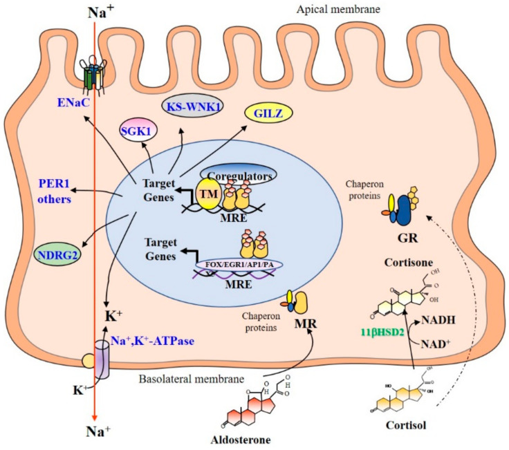
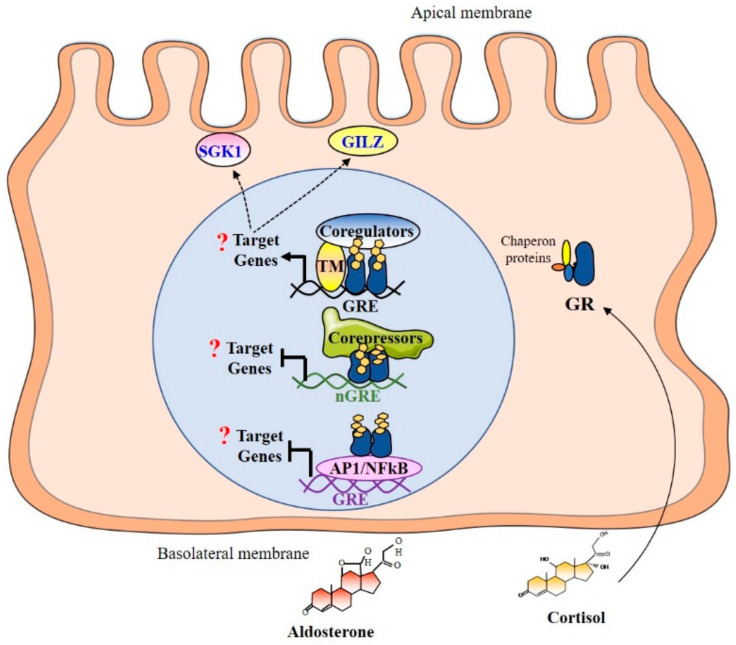
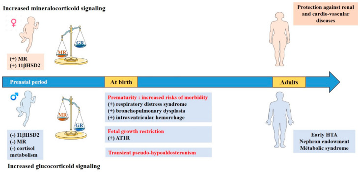
Sexual dimorphism involves differences between biological sexes that go beyond sexual characteristics. In mammals, differences between sexes have been demonstrated regarding various biological processes, including blood pressure and predisposition to develop hypertension early in adulthood, which may rely on early events during development and in the neonatal period. Recent studies suggest that corticosteroid signaling pathways (comprising glucocorticoid and mineralocorticoid signaling pathways) have distinct tissue-specific expression and regulation during this specific temporal window in a sex-dependent manner, most notably in the kidney. This review outlines the evidence for a gender differential expression and activation of renal corticosteroid signaling pathways in the mammalian fetus and neonate, from mouse to human, that may favor mineralocorticoid signaling in females and glucocorticoid signaling in males. Determining the effects of such differences may shed light on short term and long term pathophysiological consequences, markedly for males.

Keywords: aldosterone; cortisol; development; kidney; mineralocorticoid and glucocorticoid receptors; neonates; sexual dimorphism.


Conflict of interest statement

The authors declare that there is no conflict of interest that could be perceived as prejudicing the impartiality of the research.

Figures

Figure 1
Figure 1
Mineralocorticoid and glucocorticoid signaling in renal principal cells. Corticosteroid hormones enter by passive diffusion and bind their respective receptor: aldosterone to MR and cortisol/corticosterone to GR. In the absence of ligands, corticosteroid receptors are associated to chaperone proteins. Thereafter, the binding of either ligand induces the dissociation of these chaperone proteins and conformational changes of MR and GR. In the nucleus, the aldosterone/MR complex binds mostly as homodimers to Mineralocorticoid Response Elements (MREs). Then, MR interacts, in a cyclic, sequential, and/or combinatorial manner, with transcriptional coregulators and some basal transcription factors or components of the machinery to enhance the transcription of target genes, including the Epithelial Na+ Channel (ENaC), the Na+, K+-ATPase pump. Aldosterone also stimulates early expression of the Serum and glucocorticoid-regulated kinase 1 (SGK1), the serine/threonine kinase With No lysine K kinase (KS-WNK1), the N-myc Down-Regulated Gene 2 (NDRG2), and the Glucocorticoid-Induced Leucine Zipper protein (GILZ). Recently, aldosterone was shown to stimulate early expression the PER1 gene, which belongs to the circadian clock gene family. It was also reported that MR can indirectly bind to recognition motifs for other transcription factors (FOX, EGR1, AP1, PAX5) through tethering mechanisms. In principal renal cells, the 11βHSD2 converts glucocorticoid hormones into cortisone or 11-dehydrocorticosterone that have little or no affinity for MR, or even for GR. Thus, the 11βHSD2 allows aldosterone to act selectively onto MR to specifically exert its biological effects on sodium reabsorption. In addition, GR is not or weakly activated. MR: Mineralocorticoid Receptor; GR: Glucocorticoid Receptor; MRE: Mineralocorticoid Response Element; GILZ: Glucocorticoid-induced leucine zipper; ENaC: Epithelial Na+ Channel; Sgk 1: Serum and Glucocorticoid-Regulated kinase 1; KS-WNK1: With No lysine K kinase; NDRG2: N-myc Down-Regulated Gene 2; PER 1: clock gene period 1; TM: Transcriptional Machinery.
Figure 2
Figure 2
Mineralocorticoid and glucocorticoid signaling in renal principal cell during the perinatal period. MR signaling pathway is ineffective during renal development due to the down-regulation of MR expression. Given that renal 11βHSD2 is not expressed during this specific perinatal period, the GR signaling pathway is therefore functional. In the nucleus, the GR can bind specific sequences called Glucocorticoid Response Elements (GREs). Binding to the GRE activates the recruitment of chromatin-remodeling complexes and coregulators. Negative GRE (nGRE) were also reported, which are responsible for trans-repression of the target genes through the binding of GR monomers. Binding to nGRE prevents the dimerization and allows for the recruitment of corepressors, thus preventing the transcription of target genes. GR can also mediate trans-repression of target genes by tethering via interactions with other transcription factors as NFκB or AP-1 without direct binding to DNA. GR: Glucocorticoid Receptor; GRE: Glucocorticoid Response Element; GILZ: Glucocorticoid-induced leucine zipper; SGK1: Serum and Glucocorticoid-Regulated kinase 1; PER 1: clock gene period 1; TM: Transcriptional Machinery.
Figure 3
Figure 3
Sexual dimorphism of corticosteroid signaling during kidney development and consequences in pathophysiology. Sexual dimorphism of corticosteroid signaling is effective as early as the perinatal period with higher MR expression and 11βHSD2 activity in females and higher cortisol metabolism in males, thus favoring mineralocorticoid signaling in female fetus and neonates, and preferential activation of the glucocorticoid pathway in males. These imbalances may have an impact under certain pathophysiological conditions with a higher susceptibility for males to develop short and long term consequences after exacerbated activation of perinatal glucocorticoid signaling pathway.

Similar articles

Cited by

References

    1. Seccia T.M., Caroccia B., Gomez-Sanchez E.P., Gomez-Sanchez C.E., Rossi G.P. The Biology of Normal Zona Glomerulosa and Aldosterone-Producing Adenoma: Pathological Implications. Endocr. Rev. 2018;39:1029–1056. doi: 10.1210/er.2018-00060. - DOI - PMC - PubMed
    1. Miller W.L. Steroidogenesis: Unanswered Questions. Trends Endocrinol. Metab. 2017;28:771–793. doi: 10.1016/j.tem.2017.09.002. - DOI - PubMed
    1. Lefebvre H., Duparc C., Naccache A., Lopez A.-G., Castanet M., Louiset E. Paracrine Regulation of Aldosterone Secretion in Physiological and Pathophysiological Conditions. Vitam. Horm. 2019;109:303–339. doi: 10.1016/bs.vh.2018.10.001. - DOI - PubMed
    1. Wils J., Duparc C., Cailleux A.-F., Lopez A.-G., Guiheneuf C., Boutelet I., Boyer H.-G., Dubessy C., Cherifi S., Cauliez B., et al. The Neuropeptide Substance P Regulates Aldosterone Secretion in Human Adrenals. Nat. Commun. 2020;11:2673. doi: 10.1038/s41467-020-16470-8. - DOI - PMC - PubMed
    1. MacKenzie S.M., van Kralingen J.C., Davies E. Regulation of Aldosterone Secretion. Vitam. Horm. 2019;109:241–263. doi: 10.1016/bs.vh.2018.07.001. - DOI - PubMed

MeSH terms