

Generation of Synthetic Chest X-ray Images and Detection of COVID-19: A Deep Learning Based Approach
- PMID: 34069841
- PMCID: PMC8157360
- DOI: 10.3390/diagnostics11050895
Generation of Synthetic Chest X-ray Images and Detection of COVID-19: A Deep Learning Based Approach
Abstract
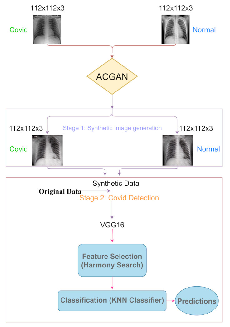
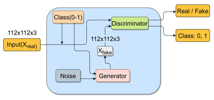
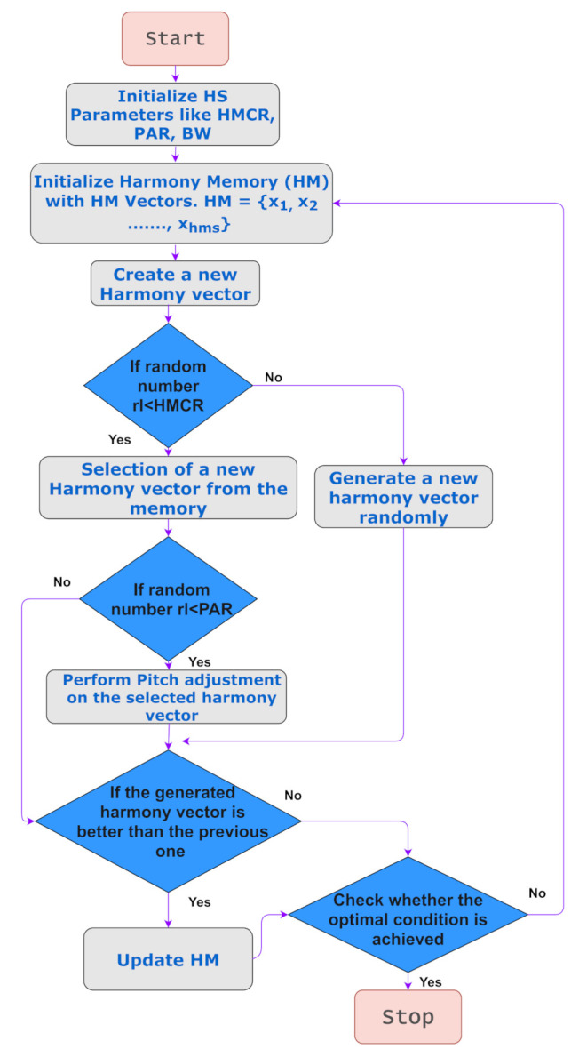
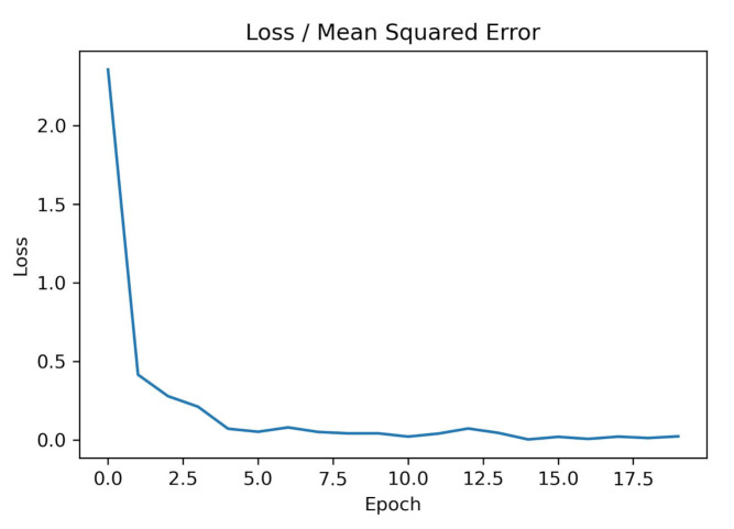
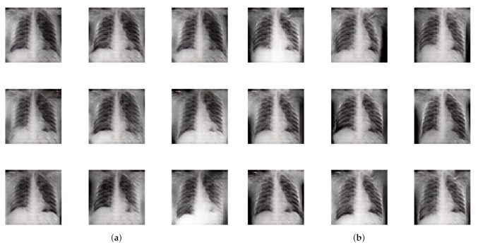
COVID-19 is a disease caused by the SARS-CoV-2 virus. The COVID-19 virus spreads when a person comes into contact with an affected individual. This is mainly through drops of saliva or nasal discharge. Most of the affected people have mild symptoms while some people develop acute respiratory distress syndrome (ARDS), which damages organs like the lungs and heart. Chest X-rays (CXRs) have been widely used to identify abnormalities that help in detecting the COVID-19 virus. They have also been used as an initial screening procedure for individuals highly suspected of being infected. However, the availability of radiographic CXRs is still scarce. This can limit the performance of deep learning (DL) based approaches for COVID-19 detection. To overcome these limitations, in this work, we developed an Auxiliary Classifier Generative Adversarial Network (ACGAN), to generate CXRs. Each generated X-ray belongs to one of the two classes COVID-19 positive or normal. To ensure the goodness of the synthetic images, we performed some experimentation on the obtained images using the latest Convolutional Neural Networks (CNNs) to detect COVID-19 in the CXRs. We fine-tuned the models and achieved more than 98% accuracy. After that, we also performed feature selection using the Harmony Search (HS) algorithm, which reduces the number of features while retaining classification accuracy. We further release a GAN-generated dataset consisting of 500 COVID-19 radiographic images.
Keywords: COVID-19 detection; chest X-ray; deep learning; feature selection; generative adversarial network; harmony search; synthetic data generation.
Conflict of interest statement
The authors declare no conflict of interest. The funders had no role in the design of the study; in the collection, analyses, or interpretation of data; in the writing of the manuscript, or in the decision to publish the results.
Figures
References
-
- Chen N., Zhou M., Dong X., Qu J., Gong F., Han Y., Qiu Y., Wang J., Liu Y., Wei Y., et al. Epidemiological and clinical characteristics of 99 cases of 2019 novel coronavirus pneumonia in Wuhan, China: A descriptive study. Lancet. 2020;395:507–513. doi: 10.1016/S0140-6736(20)30211-7. - DOI - PMC - PubMed
-
- Toussie D., Voutsinas N., Finkelstein M., Cedillo M.A., Manna S., Maron S.Z., Jacobi A., Chung M., Bernheim A., Eber C., et al. Clinical and chest radiography features determine patient outcomes in young and middle-aged adults with COVID-19. Radiology. 2020;297:E197–E206. doi: 10.1148/radiol.2020201754. - DOI - PMC - PubMed
-
- Shen B., Hoshmand-Kochi M., Abbasi A., Glass S., Jiang Z., Singer A., Thode H., Li H., Hou W., Duong T. Initial chest radiograph scores inform COVID-19 status, intensive care unit admission and need for mechanical ventilation. Clin. Radiol. 2021;76:473.e1–473.e7. doi: 10.1016/j.crad.2021.02.005. - DOI - PMC - PubMed
Grants and funding
LinkOut - more resources
Full Text Sources
Miscellaneous
