Review
. 2021 May 18;13(5):1703.
doi: 10.3390/nu13051703.

Vitamin A Update: Forms, Sources, Kinetics, Detection, Function, Deficiency, Therapeutic Use and Toxicity

Affiliations
Review

Vitamin A Update: Forms, Sources, Kinetics, Detection, Function, Deficiency, Therapeutic Use and Toxicity

Alejandro Carazo et al. Nutrients. .

Abstract

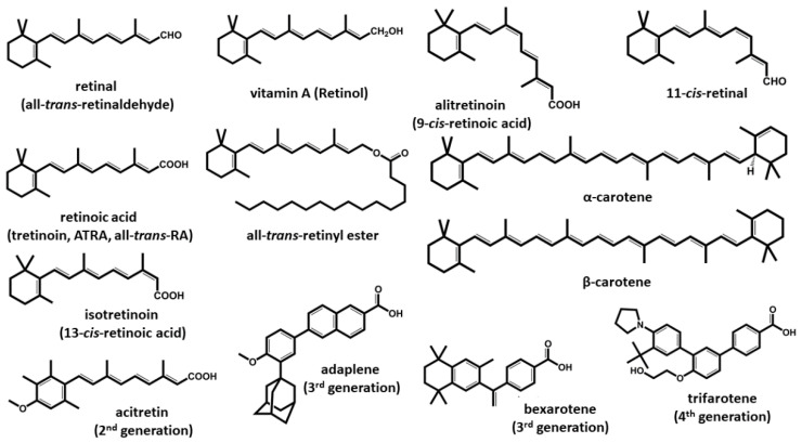
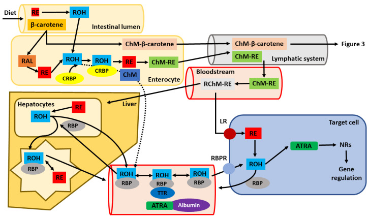
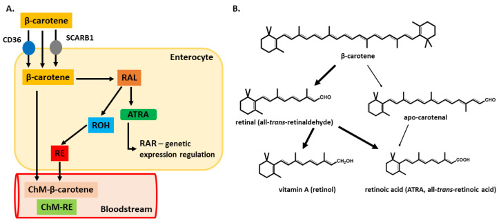
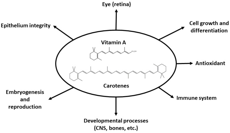
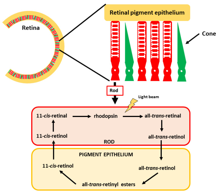
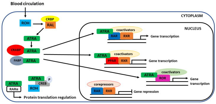
Vitamin A is a group of vital micronutrients widely present in the human diet. Animal-based products are a rich source of the retinyl ester form of the vitamin, while vegetables and fruits contain carotenoids, most of which are provitamin A. Vitamin A plays a key role in the correct functioning of multiple physiological functions. The human organism can metabolize natural forms of vitamin A and provitamin A into biologically active forms (retinol, retinal, retinoic acid), which interact with multiple molecular targets, including nuclear receptors, opsin in the retina and, according to the latest research, also some enzymes. In this review, we aim to provide a complex view on the present knowledge about vitamin A ranging from its sources through its physiological functions to consequences of its deficiency and metabolic fate up to possible pharmacological administration and potential toxicity. Current analytical methods used for its detection in real samples are included as well.

Keywords: cancer; gene regulation; hypovitaminosis; retinoic acid; retinoid receptor; retinol; toxicity; vision.


Conflict of interest statement

The authors declare no conflict of interest.

Figures

Figure 1
Figure 1
The structure of vitamin A and retinoids. The retinoids represented belong to the four described generations. First-generation compounds are found in the diet, except for some natural metabolites formed in the body. Members of the 2nd, 3rd and 4th generations are synthetic derivates based on the original retinoic structure and are used in treating different diseases. All retinoids possess a common structure and similar physicochemical properties, although their effects on the human body can vary greatly.
Figure 2
Figure 2
Cellular pathway, uptake and transport of orally given vitamin A. After metabolization in the intestinal lumen, penetration into enterocytes and its association to chylomicrons (ChM), retinyl esters (RE) and β-carotene are secreted into the lymphatic system. Later, they reach the blood (systemic circulation) and are subsequently delivered to the liver, which functions as the main retinoid storage organ in the body or target tissues/cells. The dashed line represents the portion of retinol, which is not metabolized in the intestinal cells into retinyl esters and is secreted directly into the bloodstream, where it can bind to retinol-binding protein (RBP). From the liver, retinoids can be directly secreted into the blood in association with RBP or bind later to other transport proteins (e.g., albumin) found in the blood. Transport to target tissues is enabled via the RBP-receptor (RBPR). Once they enter the target cells, retinyl esters or retinol (ROH) are further oxidized into all-trans-retinoic acid (ATRA), which is responsible for the genetic functions of vitamin A in the body (other abbreviations: RAL—retinal; RChM-RE—remnant chylomicrons-retinyl esters; TTR—transthyretin; LR—lipoprotein receptor; NRs—nuclear receptors; CRBP—cellular retinol-binding protein).
Figure 3
Figure 3
β-carotene metabolizing pathways. Absorption in the intestinal lumen can happen through passive diffusion, or it can be mediated by the membrane proteins SCARB1 and CD36. Once in the enterocyte cytoplasm, there are two possible metabolization routes. Part A shows the most common metabolization pathway y, leading finally to the secretion of retinyl esters (RE) or β-carotene into the bloodstream associated with chylomicrons. On the right (part B), both metabolic pathways are illustrated, the common route and the alternative cleavage yielding apo-carotenal molecule. Both molecules have the same metabolic end product (all-trans-RA, ATRA). The thick arrows indicate the most common pathways, while the thin arrows indicate less common metabolizing routes. Abbreviations: SCARB1—scavenger receptor class B1; RAL—retinal; ROH–retinol; CD36—cluster of differentiation 36; ChM-β—carotene–chylomicron-β-carotene; ChM-RE—chylomicron-RE).
Figure 4
Figure 4
A schematic representation of the physiological roles in which vitamin A is involved.
Figure 5
Figure 5
Vision and the role of 11-cis-retinal in the process. The retina comprises cones and rods, which mediate color and low light vision, respectively. The vitamin A derivative 11-cis-retinal is found in the rods, forming rhodopsin.
Figure 6
Figure 6
Cellular uptake by target cells and intracellular receptors for vitamin A. Once in the cytoplasm, retinol undergoes several oxidation steps, which end up forming ATRA, which can follow different fates inside the cell. ATRA can mediate both genomic and non-genomic functions. The non-genomic functions are less known and include regulation of phosphorylation of target proteins (CREB) and cytoplasmic translation regulation. Genomic functions are more common and include the binding of ATRA to nuclear receptors (RAR, PPAR, RXR, ROR), which have a direct influence on gene regulation. In the absence of a ligand, gene transcription is repressed. For this to happen, ATRA must be transported to the nucleus, which is mediated by cellular retinoic acid-binding proteins (CRABP) or fatty-acid-binding protein (FABP). CREB—cAMP response element-binding protein.

References

    1. D’Ambrosio D.N., Clugston R.D., Blaner W.S. Vitamin A metabolism: An update. Nutrients. 2011;3:63–103. doi: 10.3390/nu3010063. - DOI - PMC - PubMed
    1. Kam R.K., Deng Y., Chen Y., Zhao H. Retinoic acid synthesis and functions in early embryonic development. Cell. Biosci. 2012;2:11. doi: 10.1186/2045-3701-2-11. - DOI - PMC - PubMed
    1. Chiricozzi A., Panduri S., Dini V., Tonini A., Gualtieri B., Romanelli M. Optimizing acitretin use in patients with plaque psoriasis. Dermatol. Ther. 2017;30 doi: 10.1111/dth.12453. - DOI - PubMed
    1. Dragnev K.H., Petty W.J., Shah S.J., Lewis L.D., Black C.C., Memoli V., Nugent W.C., Hermann T., Negro-Vilar A., Rigas J.R., et al. A proof-of-principle clinical trial of bexarotene in patients with non-small cell lung cancer. Clin. Cancer Res. 2007;13:1794–1800. doi: 10.1158/1078-0432.CCR-06-1836. - DOI - PubMed
    1. Thielitz A., Gollnick H. Topical retinoids in acne vulgaris: Update on efficacy and safety. Am. J. Clin. Dermatol. 2008;9:369–381. doi: 10.2165/0128071-200809060-00003. - DOI - PubMed