Review
. 2021 May 18;10(5):1236.
doi: 10.3390/cells10051236.

Insulin Resistance and Diabetes Mellitus in Alzheimer's Disease

Affiliations
Review

Insulin Resistance and Diabetes Mellitus in Alzheimer's Disease

Jesús Burillo et al. Cells. .

Abstract

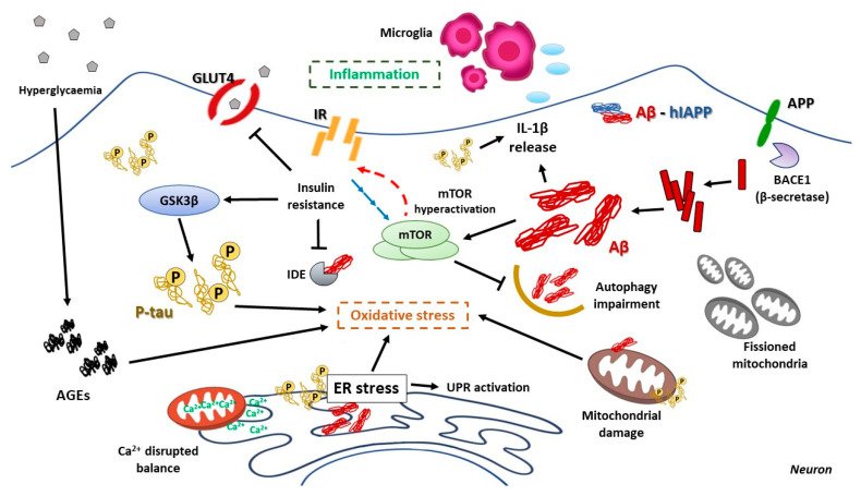
Type 2 diabetes mellitus is a progressive disease that is characterized by the appearance of insulin resistance. The term insulin resistance is very wide and could affect different proteins involved in insulin signaling, as well as other mechanisms. In this review, we have analyzed the main molecular mechanisms that could be involved in the connection between type 2 diabetes and neurodegeneration, in general, and more specifically with the appearance of Alzheimer's disease. We have studied, in more detail, the different processes involved, such as inflammation, endoplasmic reticulum stress, autophagy, and mitochondrial dysfunction.

Keywords: ER stress; T3DM; autophagy; inflammation; insulin resistance; mTOR.


Conflict of interest statement

The authors declare no conflict of interest.

Figures

Figure 1
Figure 1
Structure of mTOR complexes. Both shared mTOR that is the catalytic subunit, DEPTOR, which acts as a negative regulator, Tti1/Tel2, as scaffold proteins, and mLST8, which function is unknown. Specifically, mTORC1 is also formed with RAPTOR (scaffold protein) and PRAS40 (inhibitor of RAPTOR); mTORC2 with RICTOR and mSIN1 both acting as a scaffold; PROTOR 1/2 is a positive regulator.
Figure 2
Figure 2
Mechanisms involved in insulin and hIAPP cosecretion in pancreatic β-cell. The most important physiological stimulus of insulin secretion is glucose. Glucose is transported inside the cell thanks to GLUT2, entering the Krebs Cycle and glycolysis. These processes produced a higher ATP/ADP rate that inhibits ATP-dependent K+ channels, depolarizing the membrane. Changes in the potential of membrane open Ca2+ channels, introducing Ca2+ into the cell and promoting the release of the insulin into blood flow after glucose intake. Not only this pathway releases this hormone, but also an increment of AMPc via apetite’s hormones signaling and the activation of PKC, due to the adrenergic response. What is more important is that amylin or hIAPP is cosecreted in these insulin granules after its maturation in ER.
Figure 3
Figure 3
Molecular mechanisms of both systemic and brain insulin resistance and their consequences.
Figure 4
Figure 4
Hallmarks of pancreatic β cell failure in T2DM. Insulin secretory burden drives ER stress and misfolding of hIAPP. ER stress induces UPR activation in order to counteract protein aggregation, but chronic UPR activation leads pancreatic β-cell to apoptosis. ER stress could also promote Ca2+ unbalance among ER-mitochondria, increasing ROS and oxidative stress. Autophagy is also activated by UPR for hIAPP aggregates elimination, and its failure could promote hIAPP accumulation and detoxification via MVB-exosome secretion; pancreatic β-cell mTOR hyperactivation (due to insulin resistance or hIAPP) in turn also impaired autophagy flux. In addition, hyperglycemia and hIAPP direct interaction are thought to inhibit proteasome aggregates clearance. hIAPP mitochondrial damage could increase ROS production, oxidative stress, and accumulation of fissioned mitochondria. Hyperglycemia causes the formation of aberrant glycated molecules, the advanced glycation-end (AGEs), which increase oxidative stress. hIAPP aggregates could also induce NLRP3-induced inflammasome activation, IL-1β release, and macrophage recruitment.
Figure 5
Figure 5
Hallmarks of neuron failure in AD. Neuronal alternative processing of APP through β-secretase (BACE1) origins intracellular Aβ40-42 aggregates that will origin extracellular amyloid plaques. In parallel, microtubule-associated protein tau protein aberrant phosphorylation drives to cytoplasmic neurofibrillary tangles formation. Brain insulin resistance reduces GLUT4 glucose transporter activity and decreases insulin-degrading enzyme activity (IDE); Aβ aggregates impaired insulin and IGF-I signaling cascade. Insulin resistance results in GSK3β activation and tau hyperphosphorylation. The hyperglycaemic status also induces AGEs which are known to impair Aβ clearance and promote GSK3β-mediated tau phosphorylation. Both proteins can induce ER stress, chronic UPR activation, and neuronal apoptosis. Aβ and p-tau are known to disrupt the mitochondrial respiratory chain, increase ROS, alter Ca2+ balance and lead mitochondria to an irreversible fission status; furthermore, Parkin-mediated mitophagy is impaired in AD by Aβ and p-tau. As it happens in T2DM, insulin resistance-mediated mTOR hyperactivation affects insulin signaling, impairs neurogenesis and synaptic plasticity, and impede correct autophagy-mediated Aβ and p-tau degradation; both peptides could also affect the lysosomal function and compromise autophagosomes clearance. Aβ aggregates and p-tau tangles are known to trigger NLRP3-mediated inflammasome activation, thus releasing IL-1β and other cytokines, and inducing proinflammatory microglia recruitment. And if that were not enough, hIAPP could aggravate all these events, even worsen the situation by the generation of crossseeding heterocomplexes of Aβ-hIAPP aggregates.

Similar articles

Cited by

References

    1. Eizirik D.L., Pasquali L., Cnop M. Pancreatic β-cells in type 1 and type 2 diabetes mellitus: Different pathways to failure. Nat. Rev. Endocrinol. 2020;16:349–362. doi: 10.1038/s41574-020-0355-7. - DOI - PubMed
    1. American Diabetes Association Classification and diagnosis of diabetes: Standards of medical care in diabetes-2019. Diabetes Ca. 2019;42(Suppl. 1):S13–S28. doi: 10.2337/dc19-S002. - DOI - PubMed
    1. Pang H., Luo S., Huang G., Xia Y., Xie Z., Zhou Z. Advances in knowledge of candidate genes acting at the β-cell level in the pathogenesis of T1DM. Front. Endocrinol. 2020;11:119. doi: 10.3389/fendo.2020.00119. - DOI - PMC - PubMed
    1. Johnson M.B., Cerosaletti K., Flanagan S.E., Buckner J.H. Genetic mechanisms highlight shared pathways for the pathogenesis of polygenic type 1 diabetes and monogenic autoimmune diabetes. Curr. Diabetes Rep. 2019;19:20. doi: 10.1007/s11892-019-1141-6. - DOI - PMC - PubMed
    1. Howard S.G. Exposure to environmental chemicals and type 1 diabetes: An update. J. Epidemiol. Community Health. 2019;73:483–488. doi: 10.1136/jech-2018-210627. - DOI - PubMed

Publication types

MeSH terms