Review
. 2021 May 18;22(10):5300.
doi: 10.3390/ijms22105300.

Optogenetic Approaches for the Spatiotemporal Control of Signal Transduction Pathways

Affiliations
Review

Optogenetic Approaches for the Spatiotemporal Control of Signal Transduction Pathways

Markus M Kramer et al. Int J Mol Sci. .

Abstract

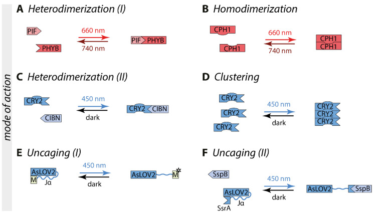
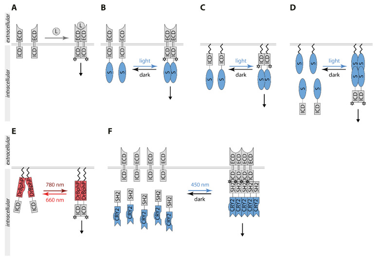
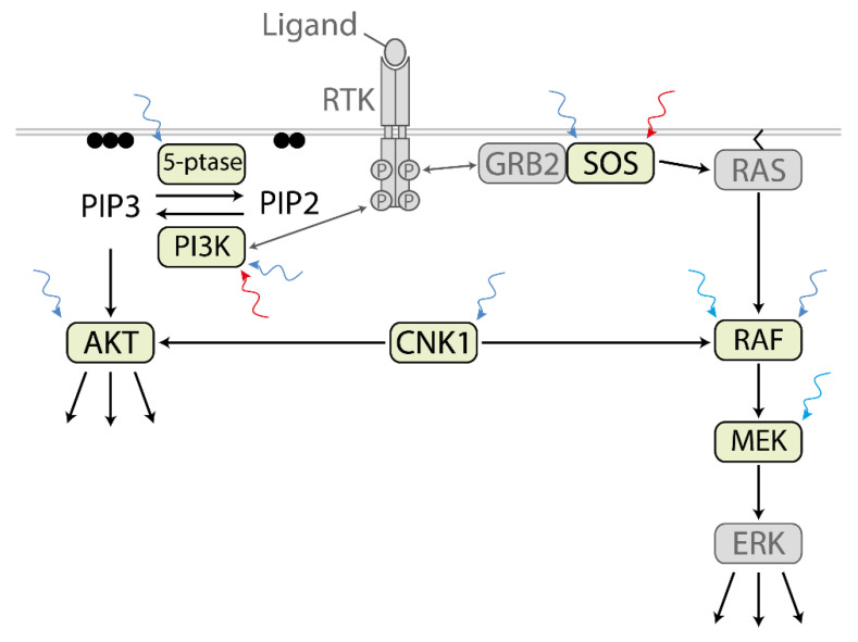
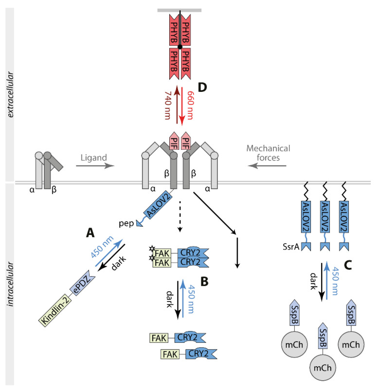
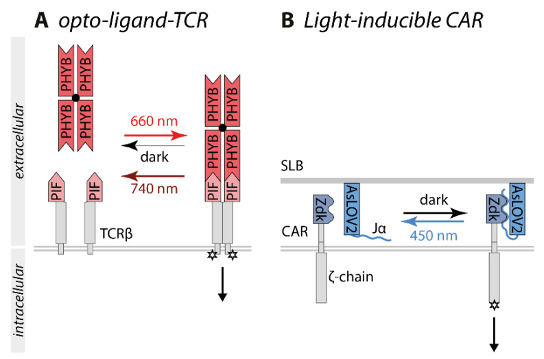
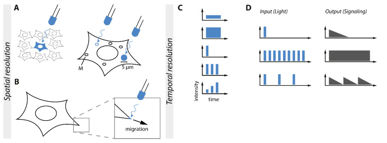
Biological signals are sensed by their respective receptors and are transduced and processed by a sophisticated intracellular signaling network leading to a signal-specific cellular response. Thereby, the response to the signal depends on the strength, the frequency, and the duration of the stimulus as well as on the subcellular signal progression. Optogenetic tools are based on genetically encoded light-sensing proteins facilitating the precise spatiotemporal control of signal transduction pathways and cell fate decisions in the absence of natural ligands. In this review, we provide an overview of optogenetic approaches connecting light-regulated protein-protein interaction or caging/uncaging events with steering the function of signaling proteins. We briefly discuss the most common optogenetic switches and their mode of action. The main part deals with the engineering and application of optogenetic tools for the control of transmembrane receptors including receptor tyrosine kinases, the T cell receptor and integrins, and their effector proteins. We also address the hallmarks of optogenetics, the spatial and temporal control of signaling events.

Keywords: control of function; optogenetics; signal transduction.


Conflict of interest statement

The authors declare no conflict of interest.

Figures

Figure 1
Figure 1
Modes of actions of optogenetic switches are illustrated on commonly used switches. (A) PHYB heterodimerizes with its interaction partner PIF upon red light illumination, which can be reversed by far-red light [18]. (B) Upon red light exposure, CPH1 forms homodimers that dissociate with far-red light [19]. (C) CRY2 engages its interaction partner CIBN to form heterodimers upon exposure with blue light [20]. (D) Additionally, CRY2 can form homo-oligomers [21]. (E) The Jα helix of AsLOV2 unwinds upon blue light illumination, thereby uncaging a motif of interest (M) [22,23]. (F) In the iLID switch, the adapter protein SsrA is exposed under blue light, enabling the binding of its interaction partner SspB [24]. Abbreviations: AsLOV2, the light-oxygen-voltage domain of phototropin 1 derived from Avena sativa; CIBN, cryptochrome-interacting basic helix-loop-helix 1 (N-terminal amino acids 1-100); CRY2, cryptochrome 2; M, the motif of interest; PHYB, phytochrome B; PIF, phytochrome interacting factor.
Figure 2
Figure 2
Schematic overview of RTK signaling controlled by optogenetic tools. (A). RTKs dimerize upon binding of the ligand (L) to the extracellular domain (ECD), stimulating the kinase activity located in the intracellular domain (ICD) and subsequently RTK downstream signaling. (B). The fusion of an optogenetic switch (S) to full-length RTKs can induce light-dependent dimerization and signal independent of ligand binding. (C). An optogenetic switch fused to the intracellular domain of RTKs can induce light-dependent dimerization of ICD and RTK signaling. (D). An optogenetic switch anchored to the plasma membrane and fused to the intracellular domain of RTKs can induce light-dependent dimerization of ICD at the plasma membrane, activating downstream signaling. (E). DrBphP fused to the ICD of RTKs can induce light-dependent dimerization of the ICD, activating downstream signaling. (F). Clustering indirectly using CRY2 (CLICR). Though clustering of CRY2 fused to a specific SH2 domain, endogenous RTKs can be targeted and activated independently of ligand stimulation. Abbreviations: CRY2, cryptochrome 2; DrBphP, Deinococcus radiodurans bacterial phytochrome; ECD, extracellular domain; ICD, intracellular domain; L, ligand; SH2, Src homology region 2.
Figure 3
Figure 3
Optogenetic control of PI3K/AKT and MAPK signaling. A wide variety of optogenetic tools is available for control of individual signaling proteins downstream of receptor tyrosine kinases (RTK). Different optogenetic switches were used for the control of the respective signaling protein. SOS: PHYB/PIF [57] and iLID [58]. RAF: CRY2/CRY2 [59], CRY2high [27], CRY2low [27] and CRY2/CIBN [60,61], pdDronpa1 [32]. MEK: pdDronpa1-based psMEK1 [32,62]. 5-ptase: CRY2/CIBN [63]. PI3K: CRY2/CIBN [64], Magnets [29], PYHB/PIF [65]. AKT: CRY2/CIBN [66,67]. CNK1: CRY2/CRY2 [68]. Abbreviations: 5-ptase, inositol 5-phosphatase OCRL; GRB2, growth factor-regulated binding protein 2; PIP2, phosphatidylinositol 4,5-bisphosphate; PIP3, phosphatidylinositol (3,4,5)-trisphosphate; RTK, receptor tyrosine kinase.
Figure 4
Figure 4
Optogenetic control of integrin signaling. (A) Using the TULIP switch the adaptor protein kindlin-2 interacts with the cytoplasmic tail of β3 inducing integrin signaling [80]. (B) Homooligomerization of focal adhesion kinase FAK by fusion to CRY2 stimulates signaling at focal adhesion sites [81]. (C) The iLID switch allows the recruitment of SspB-mCherry (mCh) to plasma membrane-anchored AsLOV2-SsrA inducing localized curvature and tension decrease correlating with stimulation of integrin signaling [82]. (D). PIF inserted in the extracellular domain of β3 mediates integrin clustering by binding matrix-bound PHYB correlating with activation of integrin signaling [81]. Abbreviations: AsLOV2, the light-oxygen-voltage domain of phototropin 1 derived from Avena sativa; CRY2, cryptochrome 2; ePDZ, engineered PDZ domain; FAK, focal adhesion kinase; mCh, mCherry; pep, peptide binding to ePDZ; PHYB, phytochrome B; PIF, phytochrome interacting factor; SspB, an interaction partner of SsrA; SsrA, adaptor protein.
Figure 5
Figure 5
Optogenetic control of T cell signaling. (A) In the opto-ligand-TCR system, PIF is inserted into the extracellular domain of TCRβ and tetramerized PHYB serves as ligand. Continuous red light exposure leads to the constant switching between the PIF-binding and non-binding states of PHYB. Higher light intensities induce higher switching rates, enabling the control of the binding half-life by the light intensity applied [87]. (B) The LOVTRAP-based Zdk CAR comprises Zdk fused to CAR and AsLOV2, presented on a supported lipid bilayer, as ligand. CAR-Zdk interacts with AsLOV2 stimulating ζ-chain-dependent signaling in the dark. Upon blue light exposure, AsLOV2 and Zdk dissociate correlating with the termination of signaling. The binding half-life can be controlled by the light intensity [88]. Abbreviations: AsLOV2, the light-oxygen-voltage domain of phototropin 1 derived from Avena sativa; CAR, chimeric antigen receptor; PHYB, phytochrome B; PIF, phytochrome interacting factor; SLB, supported lipid bilayer; TCR, T cell receptor.
Figure 6
Figure 6
Optogenetics enables unmatched spatiotemporal control of intracellular signaling. (A). Schematic overview of the spatial resolution of optogenetics. Single cells or even subcellular sections and compartments of the cell such as mitochondria (M) can be individually addressed. (B). Small sections of the membrane can be illuminated to induce migration. (C). Schematic overview of the temporal resolution of optogenetics, facilitating complex input patterns. (D). Correlation between input and output. Varying input light patterns induce different output signaling patterns generated by the same signaling module as shown for optoTGFBR [7].

Similar articles

Cited by

References

    1. Stanton B.Z., Chory E.J., Crabtree G.R. Chemically induced proximity in biology and medicine. Science. 2018;359 doi: 10.1126/science.aao5902. - DOI - PMC - PubMed
    1. Oh T.-J., Fan H., Skeeters S.S., Zhang K. Steering Molecular Activity with Optogenetics: Recent Advances and Perspectives. Adv. Biol. 2021:2000180. doi: 10.1002/adbi.202000180. - DOI - PMC - PubMed
    1. Lee H.N., Mehta S., Zhang J. Recent advances in the use of genetically encodable optical tools to elicit and monitor signaling events. Curr. Opin. Cell Biol. 2020;63:114–124. doi: 10.1016/j.ceb.2020.01.007. - DOI - PMC - PubMed
    1. Repina N.A., McClave T., Johnson H.J., Bao X., Kane R.S., Schaffer D.V. Engineered Illumination Devices for Optogenetic Control of Cellular Signaling Dynamics. Cell Rep. 2020;31:107737. doi: 10.1016/j.celrep.2020.107737. - DOI - PMC - PubMed
    1. Kramer M.M., Mühlhäuser W.W.D., Weber W., Radziwill G. Multichromatic Control of Signaling Pathways in Mammalian Cells. Adv. Biosyst. 2020:e2000196. doi: 10.1002/adbi.202000196. - DOI - PubMed

Substances

LinkOut - more resources