

Differential Effect of Non-Thermal Plasma RONS on Two Human Leukemic Cell Populations
- PMID: 34069922
- PMCID: PMC8157554
- DOI: 10.3390/cancers13102437
Differential Effect of Non-Thermal Plasma RONS on Two Human Leukemic Cell Populations
Abstract
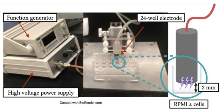
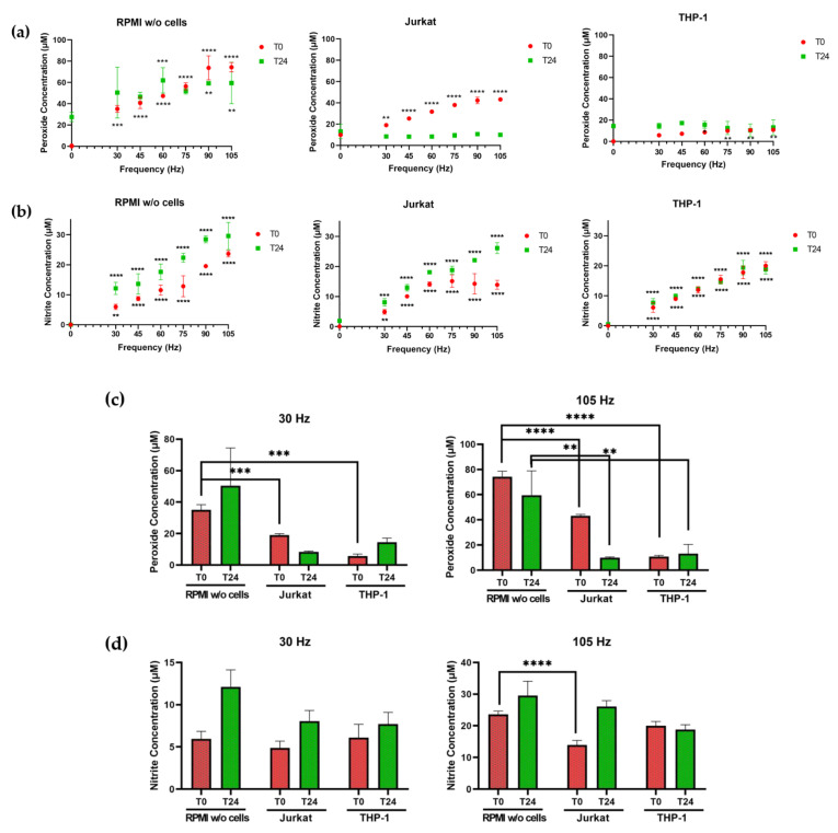
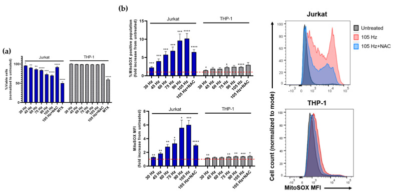
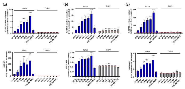
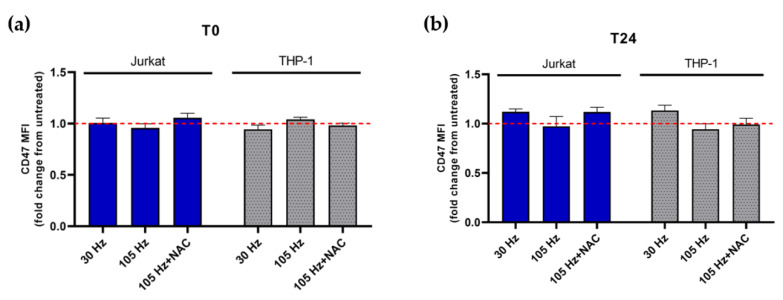
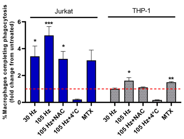
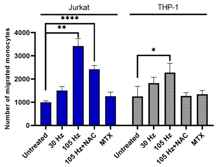
Non-thermal plasma application to cancer cells is known to induce oxidative stress, cytotoxicity and indirect immunostimulatory effects on antigen presenting cells (APCs). The purpose of this study was to evaluate the responses of two leukemic cell lines-Jurkat T lymphocytes and THP-1 monocytes-to NTP-generated reactive oxygen and nitrogen species (RONS). Both cell types depleted hydrogen peroxide, but THP-1 cells neutralized it almost immediately. Jurkat cells transiently blunted the frequency-dependent increase in nitrite concentrations in contrast to THP-1 cells, which exhibited no immediate effect. A direct relationship between frequency-dependent cytotoxicity and mitochondrial superoxide was observed only in Jurkat cells. Jurkat cells were very responsive to NTP in their display of calreticulin and heat shock proteins 70 and 90. In contrast, THP-1 cells were minimally responsive or unresponsive. Despite no NTP-dependent decrease in cell surface display of CD47 in either cell line, both cell types induced migration of and phagocytosis by APCs. Our results demonstrate that cells modulate the RONS-mediated changes in liquid chemistry, and, importantly, the resultant immunomodulatory effects of NTP can be independent of NTP-induced cytotoxicity.
Keywords: calreticulin; damage-associated molecular patterns (DAMPs); heat shock proteins; hydrogen peroxide; immunotherapy; nitrite; oxidative stress; phagocytosis; plasma medicine.
Conflict of interest statement
The authors declare no conflict of interest.
Figures
Similar articles
-
Impact of non-thermal plasma treatment on MAPK signaling pathways of human immune cell lines.Immunobiology. 2013 Oct;218(10):1248-55. doi: 10.1016/j.imbio.2013.04.015. Epub 2013 May 3. Immunobiology. 2013. PMID: 23735483
-
Non-Thermal Plasma, as a New Physicochemical Source, to Induce Redox Imbalance and Subsequent Cell Death in Liver Cancer Cell Lines.Cell Physiol Biochem. 2019;52(1):119-140. doi: 10.33594/000000009. Epub 2019 Feb 18. Cell Physiol Biochem. 2019. PMID: 30790509
-
Oxidation of Innate Immune Checkpoint CD47 on Cancer Cells with Non-Thermal Plasma.Cancers (Basel). 2021 Feb 2;13(3):579. doi: 10.3390/cancers13030579. Cancers (Basel). 2021. PMID: 33540720 Free PMC article.
-
Molecular mechanisms of non-thermal plasma-induced effects in cancer cells.Biol Chem. 2018 Dec 19;400(1):87-91. doi: 10.1515/hsz-2018-0199. Biol Chem. 2018. PMID: 30291778 Review.
-
The impact of environmental contamination on the generation of reactive oxygen and nitrogen species - Consequences for plants and humans.Environ Int. 2018 Oct;119:133-151. doi: 10.1016/j.envint.2018.06.019. Epub 2018 Jun 26. Environ Int. 2018. PMID: 29957355 Review.
Cited by
-
Current State of Cold Atmospheric Plasma and Cancer-Immunity Cycle: Therapeutic Relevance and Overcoming Clinical Limitations Using Hydrogels.Adv Sci (Weinh). 2023 Mar;10(8):e2205803. doi: 10.1002/advs.202205803. Epub 2023 Jan 20. Adv Sci (Weinh). 2023. PMID: 36670068 Free PMC article. Review.
-
Human head and neck cancer cell lines response to cold atmospheric plasma activated media is affected by the chemistry of culture media.Heliyon. 2024 Dec 25;11(1):e41458. doi: 10.1016/j.heliyon.2024.e41458. eCollection 2025 Jan 15. Heliyon. 2024. PMID: 39866438 Free PMC article.
-
Reactive oxygen species from non-thermal gas plasma (CAP): implication for targeting cancer stem cells.Cancer Cell Int. 2024 Oct 22;24(1):344. doi: 10.1186/s12935-024-03523-x. Cancer Cell Int. 2024. PMID: 39438918 Free PMC article. Review.
-
Immunomodulatory Effects of Non-Thermal Plasma in a Model for Latent HIV-1 Infection: Implications for an HIV-1-Specific Immunotherapy.Biomedicines. 2023 Jan 3;11(1):122. doi: 10.3390/biomedicines11010122. Biomedicines. 2023. PMID: 36672628 Free PMC article.
-
Effect of plasma-induced oxidation on NK cell immune checkpoint ligands: A computational-experimental approach.Redox Biol. 2024 Nov;77:103381. doi: 10.1016/j.redox.2024.103381. Epub 2024 Oct 1. Redox Biol. 2024. PMID: 39395241 Free PMC article.
References
-
- Semmler M.L., Bekeschus S., Schäfer M., Bernhardt T., Fischer T., Witzke K., Seebauer C., Rebl H., Grambow E., Vollmar B., et al. Molecular Mechanisms of the Efficacy of Cold Atmospheric Pressure Plasma (CAP) in Cancer Treatment. Cancers. 2020;12:269. doi: 10.3390/cancers12020269. - DOI - PMC - PubMed
-
- Liedtke K.R., Bekeschus S., Kaeding A., Hackbarth C., Kuehn J.P., Heidecke C.D., von Bernstorff W., von Woedtke T., Partecke L.I. Non-thermal plasma-treated solution demonstrates antitumor activity against pancreatic cancer cells in vitro and in vivo. Sci. Rep. 2017;7:8319. doi: 10.1038/s41598-017-08560-3. - DOI - PMC - PubMed
-
- Lin A., Gorbanev Y., De Backer J., Van Loenhout J., Van Boxem W., Lemière F., Cos P., Dewilde S., Smits E., Bogaerts A. Non-Thermal Plasma as a Unique Delivery System of Short-Lived Reactive Oxygen and Nitrogen Species for Immunogenic Cell Death in Melanoma Cells. Adv. Sci. 2019;6:1802062. doi: 10.1002/advs.201802062. - DOI - PMC - PubMed
LinkOut - more resources
Full Text Sources
Research Materials
Miscellaneous
