. 2021 May 18;11(5):1330.
doi: 10.3390/nano11051330.

Cytokine-Mediated Inflammation in the Oral Cavity and Its Effect on Lipid Nanocarriers

Affiliations

Cytokine-Mediated Inflammation in the Oral Cavity and Its Effect on Lipid Nanocarriers

Carolin Tetyczka et al. Nanomaterials (Basel). .

Abstract

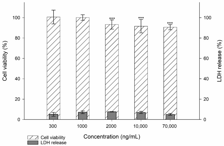
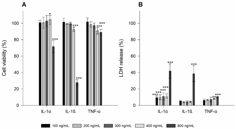
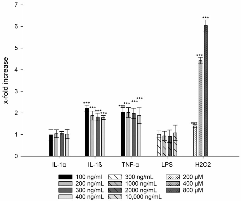
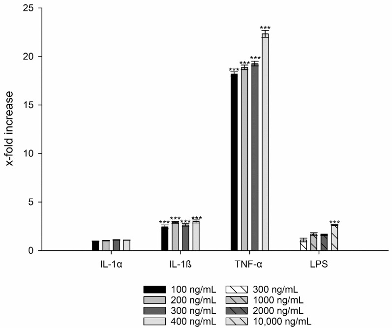
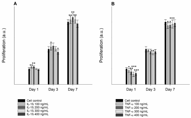
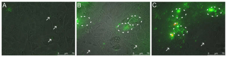
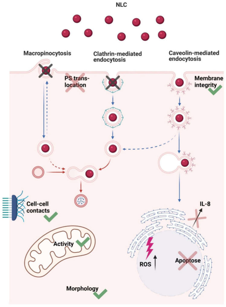
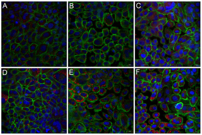
Topical drug administration to the oral mucosa proves to be a promising treatment alternative for inflammatory diseases. However, disease-related changes in the cell barrier must be considered when developing such delivery systems. This study aimed at investigating the changes in the lining mucosa caused by inflammation and evaluating the consequences on drug delivery systems such as nanostructured lipid carriers (NLC). For this, TR146 cells were treated with inflammatory cytokines and bacterial components. Cell viability and integrity, reactive oxygen species (ROS), and interleukin (IL)-8 release were used as endpoints to assess inflammation. Translocation of phosphatidylserine, cytoskeletal arrangement, opening of desmosomes, and cell proliferation were examined. Transport studies with NLC were performed considering active and passive pathways. The results showed that IL-1ß and tumor necrosis factor α induced inflammation by increasing IL-8 and ROS production (22-fold and 2-fold). Morphologically, loss of cell-cell connections and formation of stress fibers and hyperplasia were observed. The charge of the cell membrane shifted from neutral to negative, which increased the absorption of NLC due to the repulsive interactions between the hydrophobic negative particles and the cell membrane on the one hand, and interactions with lipophilic membrane proteins such as caveolin on the other.

Keywords: TR146; cytokine; in-vitro inflammation; lining mucosa; nanostructured lipid carriers.


Conflict of interest statement

The authors declare no conflict of interest.

Figures

Figure 1
Figure 1
Cell viability and membrane integrity (i.e., LDH release) of TR146 cells treated with 300–70,000 ng/mL LPS. The percentages given (% ± SD) refer to the corresponding controls. Student’s t-test was used for statistical analysis.Significant differences compared to the control are marked as ***, which corresponds to a p value ≤ 0.001.
Figure 2
Figure 2
Cell viability (A) and membrane integrity (i.e., LDH release; (B)) of TR146 cells treated with 100–800 ng/mL IL-1α, IL-1ß, and TNF-α. The percentages given (% ± SD) refer to the corresponding controls. Student’s t-test was used for statistical analysis. Significant differences compared to the control are marked as *, which correspond to a p value ≤ 0.05, **, which corresponds to a p value ≤ 0.01 and ***, which corresponds to a p value ≤ 0.001.
Figure 3
Figure 3
Generation of ROS after treatment with IL-1α, IL-1ß, LPS, and TNF-α compared to untreated cells. H2O2 was used as positive control. Student´s t-test was used for statistical analysis. Significant differences compared to the control are marked as ***, which corresponds to a p value ≤ 0.001.
Figure 4
Figure 4
IL-8 release of TR146 cells after treatment with LPS, IL-1α, IL-1ß, and TNF-α compared to untreated cells. Student´s t-test was used for statistical analysis. Significant differences compared to the control are marked as ***, which corresponds to a p value ≤ 0.001.
Figure 5
Figure 5
Proliferation of untreated TR146 cells and cells treated with 100–400 ng/mL IL-1ß (A) and TNF-α (B) over a period of 1, 3, and 7 days using a CellTiter 96 Aqueous Non-Radioactive Cell Proliferation Assay. Student´s t-test was used for statistical analysis. Significant differences compared to the control are marked as *, which corresponds to a p value ≤ 0.05, **, which corresponds to a p value ≤ 0.01 and ***, which corresponds to a p value ≤ 0.001.
Figure 6
Figure 6
Fluorescence microscopic images (overlay of bright field and fluorescence channels) of untreated TR146 cells (A) and cells treated with 400 ng/mL IL-1ß (B) and 300 ng/mL TNF-α (C). Circles indicate specific binding of Annexin V-Alexa Fluor 488 (green) to PS on the outside of the cells. Arrows show cell blebbing (green spots) and stars indicate PI stained necrotic cells (red).
Figure 7
Figure 7
Fluorescence microscopic images of untreated TR146 cells (A) and TR146 cells treated with 400 ng/mL IL-1ß (B) and 300 ng/mL TNF-α (C). Cell nuclei were stained with Hoechst (blue) and the cytoskeleton was stained with Alexa Fluor 488 Phalloidin (green). Arrows show the formation of stress fibers.
Figure 8
Figure 8
Schematic illustration describing NLC–cell interactions and uptake mechanism. Briefly, NLC were transported into the cell via caveolin-mediated endocytosis. Membrane integrity and mitochondrial activity were not affected; there was no translocation of PS, cell morphology was comparable to the control, and cell–cell-contacts remained intact. Apoptosis was not induced, there was no increase in IL-8 release, but ROS production was increased.
Figure 9
Figure 9
Fluorescence microscopic images of untreated TR146 cells (A,D) and TR146 cells treated with 400 ng/mL IL-1ß (B,E) and 300 ng/mL TNF-α (C,F). Particle uptake was performed with 500 µg/mL (AC) and 750 µg/mL (DF) NLC. Cell nuclei were stained with Hoechst (blue) and the cytoskeleton was stained with Alexa Fluor 488 Phalloidin (green). NLC were labeled with oil red o (red).
Figure 10
Figure 10
Fluorescence microscopic z-scans of untreated TR146 cells (A,D) and TR146 cells treated with 400 ng/mL IL-1ß (B,E) and 300 ng/mL TNF-α (C,F). Particle uptake was performed with 500 µg/mL (AC) and 750 µg/mL (DF) NLC. Cell nuclei were stained with Hoechst (blue) and the cytoskeleton was stained with Alexa Fluor 488 Phalloidin (green). The circles represent particle uptake/internalization into TR146 cells. NLC were labeled with oil red o (red).
Figure 11
Figure 11
Fluorescence microscopic z-scans of untreated TR146 cells at 4 °C (A) and TR146 cells treated with 400 ng/mL IL-1ß and 300 ng/mL TNF-α at 4 °C (B,C). TR146 cells treated with Dynasore (D) and additionally with IL-1ß and TNF-α (E,F). TR146 cells treated with Genistein (G) and additionally with IL-1ß and TNF-α (H,I). TR146 cells treated with Chlorpromazine (J) and additionally with IL-1ß and TNF-α (K,L). TR146 cells treated with EIPA (M) and additionally with IL-1ß and TNF-α (N,O). Cell nuclei were stained with Hoechst (blue) and the cytoskeleton was stained with Alexa Fluor 488 Phalloidin (green). The circles represent particle uptake/internalization into TR146 cells. NLC were labelled with oil red o (red).

Similar articles

Cited by

References

    1. World Health Organization Oral Health. [(accessed on 6 November 2019)]; Available online: https://www.who.int/news-room/fact-sheets/detail/oral-health.
    1. Platform for Better Oral Health in Europe The State of Oral Health in Europe. [(accessed on 6 November 2019)]; Available online: http://www.oralhealthplatform.eu/our-work/the-state-of-oral-health-in-eu...
    1. Hasturk H., Kantarci A., Van Dyke T.E. Oral inflammatory diseases and systemic inflammation: Role of the macrophage. Front. Immunol. 2012;3:118. doi: 10.3389/fimmu.2012.00118. - DOI - PMC - PubMed
    1. Politis C., Schoenaers J., Jacobs R., Agbaje J.O. Wound Healing Problems in the Mouth. Front. Physiol. 2016;7:507. doi: 10.3389/fphys.2016.00507. - DOI - PMC - PubMed
    1. De Oliveira S., Rosowski E.E., Huttenlocher A. Neutrophil migration in infection and wound repair: Going forward in reverse. Nat. Rev. Immunol. 2016;16:378–391. doi: 10.1038/nri.2016.49. - DOI - PMC - PubMed

LinkOut - more resources