

Impacts of the Type I Toxin-Antitoxin System, SprG1/SprF1, on Staphylococcus aureus Gene Expression
- PMID: 34070083
- PMCID: PMC8158120
- DOI: 10.3390/genes12050770
Impacts of the Type I Toxin-Antitoxin System, SprG1/SprF1, on Staphylococcus aureus Gene Expression
Abstract
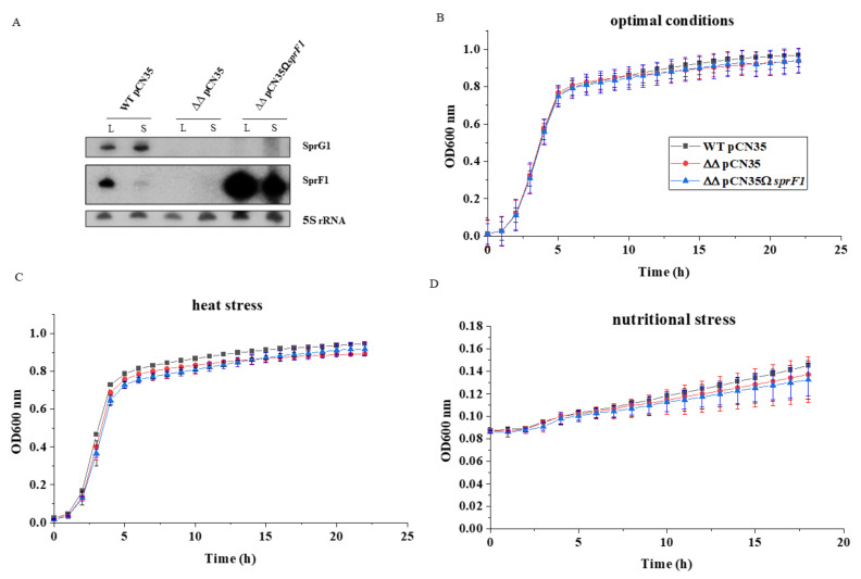
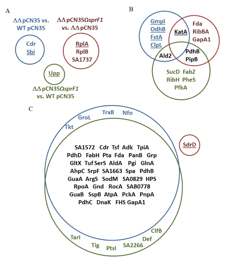
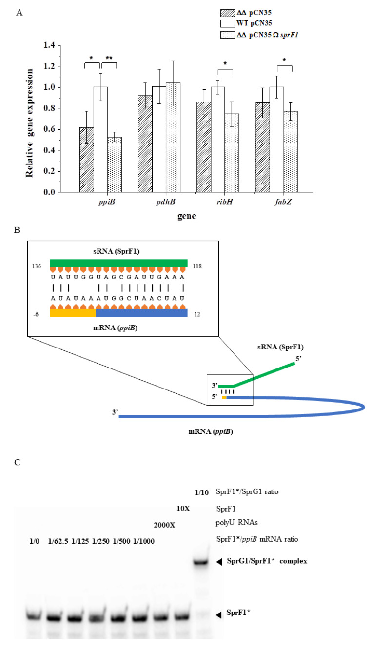
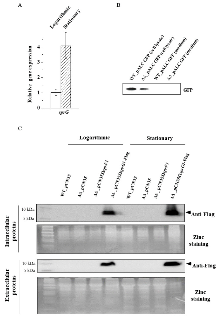
Type I toxin-antitoxin (TA) systems are widespread genetic modules in bacterial genomes. They express toxic peptides whose overexpression leads to growth arrest or cell death, whereas antitoxins regulate the expression of toxins, acting as labile antisense RNAs. The Staphylococcus aureus (S. aureus) genome contains and expresses several functional type I TA systems, but their biological functions remain unclear. Here, we addressed and challenged experimentally, by proteomics, if the type I TA system, the SprG1/SprF1 pair, influences the overall gene expression in S. aureus. Deleted and complemented S. aureus strains were analyzed for their proteomes, both intracellular and extracellular, during growth. Comparison of intracellular proteomes among the strains points to the SprF1 antitoxin as moderately downregulating protein expression. In the strain naturally expressing the SprG1 toxin, cytoplasmic proteins are excreted into the medium, but this is not due to unspecific cell leakages. Such a toxin-driven release of the cytoplasmic proteins may modulate the host inflammatory response that, in turn, could amplify the S. aureus infection spread.
Keywords: 2D-DIGE; RNA antitoxin; Staphylococcus aureus; peptide toxins; proteomics; toxin–antitoxin systems (type I).
Conflict of interest statement
The authors declare no conflict of interest. The funders had no role in the design of the study; in the collection, analyses, or interpretation of data; in the writing of the manuscript, or in the decision to publish the results.
Figures
Similar articles
-
Functionality and cross-regulation of the four SprG/SprF type I toxin-antitoxin systems in Staphylococcus aureus.Nucleic Acids Res. 2019 Feb 28;47(4):1740-1758. doi: 10.1093/nar/gky1256. Nucleic Acids Res. 2019. PMID: 30551143 Free PMC article.
-
Bacterial Type I Toxins: Folding and Membrane Interactions.Toxins (Basel). 2021 Jul 14;13(7):490. doi: 10.3390/toxins13070490. Toxins (Basel). 2021. PMID: 34357962 Free PMC article. Review.
-
Genome-Wide Screening for Identification of Novel Toxin-Antitoxin Systems in Staphylococcus aureus.Appl Environ Microbiol. 2019 Oct 1;85(20):e00915-19. doi: 10.1128/AEM.00915-19. Print 2019 Oct 15. Appl Environ Microbiol. 2019. PMID: 31375497 Free PMC article.
-
Toxin-Antitoxin Systems of Staphylococcus aureus.Toxins (Basel). 2016 May 5;8(5):140. doi: 10.3390/toxins8050140. Toxins (Basel). 2016. PMID: 27164142 Free PMC article. Review.
-
A novel Staphylococcus aureus cis-trans type I toxin-antitoxin module with dual effects on bacteria and host cells.Nucleic Acids Res. 2019 Feb 28;47(4):1759-1773. doi: 10.1093/nar/gky1257. Nucleic Acids Res. 2019. PMID: 30544243 Free PMC article.
Cited by
-
Differential Virulence of Aggregatibacter actinomycetemcomitans Serotypes Explained by Exoproteome Heterogeneity.Microbiol Spectr. 2023 Feb 14;11(1):e0329822. doi: 10.1128/spectrum.03298-22. Epub 2022 Dec 21. Microbiol Spectr. 2023. PMID: 36541765 Free PMC article.
-
Thirty Years of sRNA-Mediated Regulation in Staphylococcus aureus: From Initial Discoveries to In Vivo Biological Implications.Int J Mol Sci. 2022 Jul 1;23(13):7346. doi: 10.3390/ijms23137346. Int J Mol Sci. 2022. PMID: 35806357 Free PMC article. Review.
References
-
- Zamakhaev M.V., Goncharenko A.V., Shumkov M.S. Toxin-Antitoxin Systems and Bacterial Persistence (Review) Appl. Biochem. Microbiol. 2019;55:571–581. doi: 10.1134/S0003683819060140. - DOI
Publication types
MeSH terms
Substances
LinkOut - more resources
Full Text Sources
Molecular Biology Databases
