. 2021 May 18;9(5):569.
doi: 10.3390/biomedicines9050569.

Influence of Serotonin 5-HT4 Receptors on Responses to Cardiac Stressors in Transgenic Mouse Models

Affiliations

Influence of Serotonin 5-HT4 Receptors on Responses to Cardiac Stressors in Transgenic Mouse Models

Ulrich Gergs et al. Biomedicines. .

Abstract

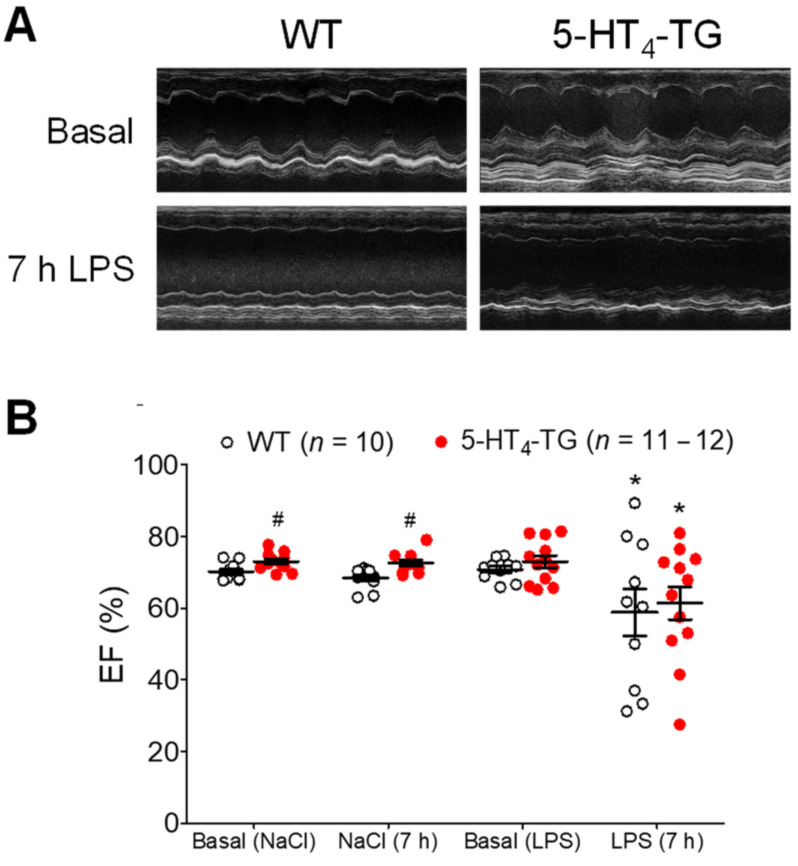
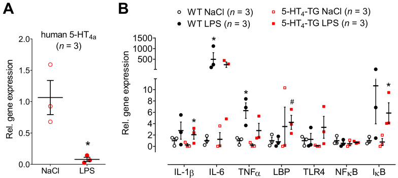
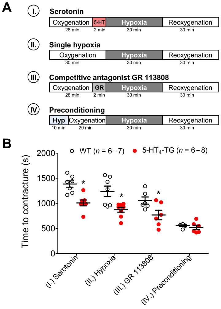
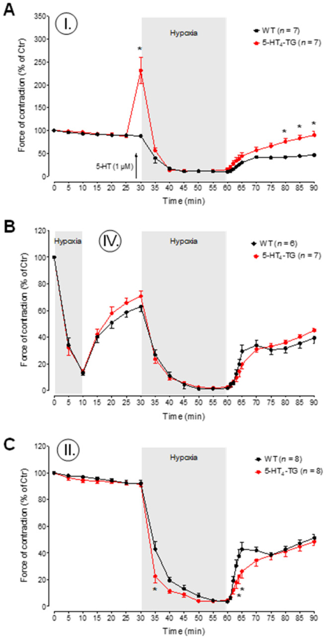
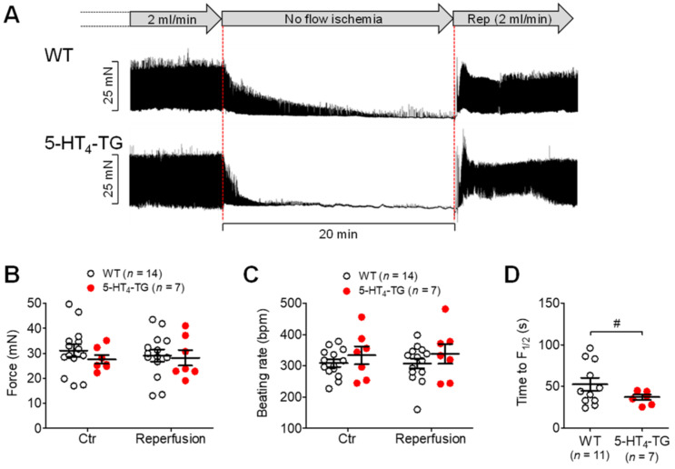
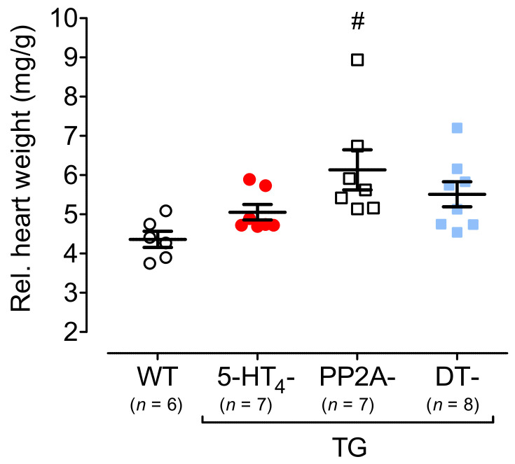
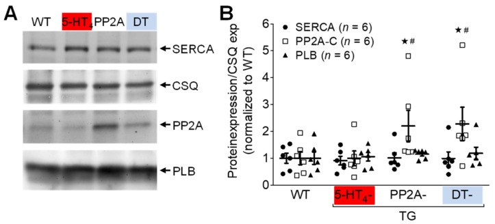
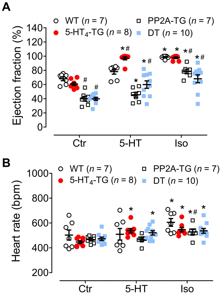
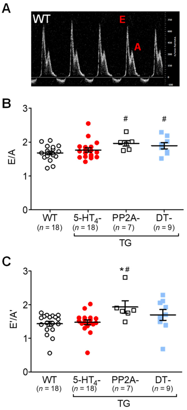
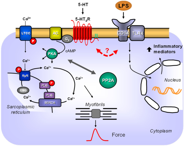
The current study aimed to deepen our knowledge on the role of cardiac 5-HT4 receptors under pathophysiological conditions. To this end, we used transgenic (TG) mice that overexpressed human 5-HT4a receptors solely in cardiac myocytes (5-HT4-TG mice) and their wild-type (WT) littermates that do not have functional cardiac 5-HT4 receptors as controls. We found that an inflammation induced by lipopolysaccharide (LPS) was detrimental to cardiac function in both 5-HT4-TG and WT mice. In a hypoxia model, isolated left atrial preparations from the 5-HT4-TG mice went into contracture faster during hypoxia and recovered slower following hypoxia than the WT mice. Similarly, using isolated perfused hearts, 5-HT4-TG mice hearts were more susceptible to ischemia compared to WT hearts. To study the influence of 5-HT4 receptors on cardiac hypertrophy, 5-HT4-TG mice were crossbred with TG mice overexpressing the catalytic subunit of PP2A in cardiac myocytes (PP2A-TG mice, a model for genetically induced hypertrophy). The cardiac contractility, determined by echocardiography, of the resulting double transgenic mice was attenuated like in the mono-transgenic PP2A-TG and, therefore, largely determined by the overexpression of PP2A. In summary, depending on the kind of stress put upon the animal or isolated tissue, 5-HT4 receptor overexpression could be either neutral (genetically induced hypertrophy, sepsis) or possibly detrimental (hypoxia, ischemia) for mechanical function. We suggest that depending on the underlying pathology, the activation or blockade of 5-HT4 receptors might offer novel drug therapy options in patients.

Keywords: 5-HT4 receptor; LPS; PP2A transgenic mice; cardiac hypertrophy; hypoxia; inflammation; ischemia; serotonin; transgenic mice.


Conflict of interest statement

The authors declare no conflict of interest.

Figures

Figure 1
Figure 1
Echocardiography of LPS-treated mice. (A) M-mode pictures of WT and 5-HT4-TG, basal and 7 h after LPS treatment. (B) LPS treatment (7 h) led to a deterioration of cardiac function demonstrated as decreased left ventricular ejection fraction (EF). Number in brackets indicates the number of mice studied. WT = wild-type mice, 5-HT4-TG=5-HT4-transgenic mice. Data shown are means ± SEM. * p < 0.05 vs. basal; # p < 0.05 vs. WT.
Figure 2
Figure 2
mRNA expression in WT and 5-HT4-TG mice, treated either with LPS or NaCl. (A) The mRNA coding for the overexpressed human 5-HT4-receptor was greatly downregulated in hearts of TG after LPS treatment. Ordinate: mRNA expression normalized to GAPDH expression. Three mice were studied in each genotype. * p < 0.05 vs. NaCl. (B) LPS-induced heart failure was accompanied by increased mRNA expression of cytokines like interleukin 1 β and 6 (IL-1 β, IL-6) and tumor necrosis factor α (TNFα) in both 5-HT4-TG and WT. The mRNA of the LPS-binding protein (LBP) and the Toll-like receptor 4 (TLR4) was increased in 5-HT4-TG, but not in WT. Whereas the mRNA of NFκB was unchanged, the mRNA of IκBα was to a similar extent increased by LPS in WT and 5-HT4-TG. Three mice were studied in each group, and injection of NaCl served as control. WT = wild-type mice, 5-HT4-TG = 5-HT4-transgenic mice. Data shown are means ± SEM. * p < 0.05 vs. NaCl; # p < 0.05 vs. WT.
Figure 3
Figure 3
Hypoxia in atrial preparations. (A) The scheme demonstrates the experimental protocols of the experiments. Paced left atrial preparations from wild-type mice (WT), or 5-HT4-transgenic mice (5-HT4-TG) were allowed to equilibrate in the organ bath in buffer saturated with carbogen (=oxygenation, 5% CO2 and 95% O2). Then as indicated, four protocols were performed: (I.) 28 min of oxygenation followed by addition of serotonin (5-HT, 1 µM) for 2 min; (II.) 30 min of oxygenation; (III.) 28 min of oxygenation followed by addition of the 5-HT4-antagonist GR 113808 (GR, 1 µM) for 2 min; (IV.) 10 min of hypoxia (Hyp) followed by 20 min of oxygenation. Thereafter, all conditions (I.–IV.) include the same procedure: 30 min of hypoxia (5% CO2 and 95% N2) and then again carbogen (reoxygenation). (B) During hypoxia, left atrial preparations lose their ability to completely relax, and an increase in diastolic tension (contractures) occurs. Under the setups serotonin (1 µM), single hypoxia and GR 113808 (1 µM), 5-HT4-TG atria developed contractures earlier than WT atria. Roman numbers indicate the experimental protocol, and numbers at the bottom of the columns indicate the number of experiments. Data shown are means ± SEM. * p < 0.05 vs. WT.
Figure 4
Figure 4
Time course of hypoxia in left atrial preparations. (AC) The force of contraction in% of control (Ctr = initial force at the beginning of the experiment) during the time of oxygenation and hypoxia is presented. After the addition of serotonin (5-HT, 1 μM, protocol I), the relative force was greatly increased in 5-HT4-TG and reached initial values again after hypoxia and reoxygenation (A). Preconditioning (protocol IV) as short hypoxia for 10 min was not beneficial (B). Under the condition of single hypoxia (protocol II), force decline was faster in TG left atria than in WT (C). WT = wild-type mice, 5-HT4-TG=5-HT4-transgenic mice. Data shown are means ± SEM. * p < 0.05 vs. WT.
Figure 5
Figure 5
Basal characteristics of isolated perfused heart preparations from WT and 5-HT4-TG mice. (A) Basal force of contraction in mN. (B) Effects of agonists and antagonists on the force of contraction after 5 min (maximum was reached). (C) Basal beating rate in beats per minute (bpm). (D) Effects of agonists and antagonists on the beating rate after 5 min. Ctr, control, Iso, isoproterenol (1 µM), 5-HT, serotonin (1 µM) and 5-HT (1 µM) in the presence of the 5-HT4-receptor antagonist’s GR 113808 (1 µM) or GR 125487 (1 µM). WT = wild-type mice, 5-HT4-TG = 5-HT4-transgenic mice. Data shown are means ± SEM. * p < 0.05 vs. Ctr, # p < 0.05 vs. WT.
Figure 6
Figure 6
Ischemia and reperfusion in isolated perfused heart preparations. (A) Exemplary recordings of the time course of force reduction (ischemia) and force recovery (reperfusion, Rep) in isolated perfused heart preparations from WT and 5-HT4-TG. The perfusion rate was always 2 mL/min. No flow ischemia indicates global ischemia of the heart by stopping the perfusion pump. Horizontal bar: 20 min of ischemia. A period of 20 min ischemia did not cause permanent damage because, after reperfusion, force (B) and heart rate (C) of both 5-HT4-TG and WT reached preischemic values again. Time to 50% decline of developed force (F½) during ischemia was reduced in 5-HT4-TG compared to WT (D). WT = wild-type mice, 5-HT4-TG=5-HT4-transgenic mice. Data shown are means ± SEM. # p < 0.05 vs. WT.
Figure 7
Figure 7
Protein phosphorylation after ischemia/reperfusion in isolated perfused hearts of WT and 5-HT4-TG mice. (A) The scheme demonstrates the protocols (Langendorff perfusion: 2 mL/min flow): (1) 15 min equilibration, 20 min ischemia by stopping the perfusion followed by 15 min reperfusion or 50 min continuous perfusion with saline buffer as time control; (2) 15 min equilibration, 20 min ischemia and 15 min reperfusion in the presence of 1 µM serotonin (5-HT) or 35 min perfusion followed by 15 min perfusion with 5-HT (1 µM) as time control without ischemia. (B) Representative Western blots. The loading scheme is shown in the table above the blots. TG = 5-HT4-TG. (C) Phosphorylation of phospholamban at serine-16 (PS16-PLB) and (D) threonine-17 (PT17-PLB) normalized to cardiac calsequestrin (CSQ). (E) Phosphorylation of the mitogen-activated protein kinases (MAPK) p38 and (F) ERK1/2 normalized to the non-phosphorylated MAPKs. Ordinates: Ratio of phosphoproteins to calsequestrin or non-phosphorylated MAPKs in arbitrary imager units. Data shown are means ± SEM. * p < 0.05 vs. Ctr; § p < 0.05 vs. ischemia; + p < 0.05 vs. Ctr + 5-HT. WT = wild-type mice, 5-HT4-TG = 5-HT4-transgenic mice; n.d., not determined (As WT preparations did not respond to 5-HT, perfusion with 5-HT was exclusively done with 5-HT4-TG hearts).
Figure 8
Figure 8
Heart weight. Relative heart weights of 5-HT4-TG, PP2A-TG and double transgenic (DT) mice at 12 months of age compared to wild-type (WT) mice. Ordinate: heart weight in milligrams (mg) divided by body weight in grams (g). TG, transgenic mice. Numbers in brackets indicate the numbers of mice studied. Data shown are means ± SEM. # p < 0.05 vs. WT.
Figure 9
Figure 9
Protein expression in double transgenic mice. Protein expression of SERCA, PP2A, PLB and CSQ in hearts of wild-type (WT), 5-HT4-TG, PP2A-TG and double transgenic (DT) mice. (A) Representative Western blots. (B) Quantification of ventricular proteins. Data were normalized to CSQ (loading control) and to mean WT expression. TG, transgenic mice. Data shown are means ± SEM. # p < 0.05 vs. WT;  p < 0.05 vs. 5-HT4-TG.
Figure 10
Figure 10
Echocardiography of double transgenic mice. Echocardiography of wild-type (WT), 5-HT4-transgenic (5-HT4-TG), PP2A-transgenic (PP2A-TG) and double transgenic (DT) mice. (A) Basal ejection fraction (Ctr) was reduced in PP2A-TG and DT mice, and 5-HT increased EF only in 5-HT4-TG and DT mice. β-adrenergic stimulation by isoproterenol (Iso) increased EF less in PP2A-TG and DT compared to the other groups. (B) Basal heart rate (Ctr) was not different between genotypes, and positive chronotropic effects of 5-HT were only noted in 5-HT4-TG and DT mice. However, β-adrenergic stimulation (Iso) increased heart rate in all groups. Numbers in bars indicate the numbers of mice studied. Data shown are means ± SEM. * p < 0.05 vs. Ctr; # p < 0.05 vs. WT.
Figure 11
Figure 11
Doppler echocardiography of double transgenic mice. Pulsed wave (PW) Doppler echocardiography of wild-type (WT), 5-HT4-transgenic (5-HT4-TG), PP2A-transgenic (PP2A-TG) and double transgenic (DT) mice. (A) A typical pattern of E wave and A wave in mitral flow. The E wave represents the early filling of the ventricle. The A wave represents the atrial contraction. (B) E divided by A was increased in PP2A-TG and in DT. (C) By tissue Doppler imaging of the left ventricular posterior wall, the early (E’) and late (A’) diastolic and systolic maximum tissue velocity was assessed. The E’ wave corresponds to the motion of the posterior wall during early diastolic filling of the left ventricle, and the A’ wave originates from atrial contraction during the late filling of the left ventricle. An increased E’/A’ quotient was noted in PP2A-TG but not in DT mice. Numbers in bars indicate the numbers of mice studied. Data shown are means ± SEM. # p < 0.05 vs. WT;  p < 0.05 vs. 5-HT4-TG.
Figure 12
Figure 12
Scheme. 5-HT signaling via 5-HT4-receptors and LPS signaling in TG cardiac myocytes. Stimulation of cardiac 5-HT4-receptors in the sarcolemma of transgenic mice leads to stimulation of adenylate cyclase (AC) via stimulatory G-proteins (Gs). AC increases cAMP levels in the cytosol, where it can either directly activate HCN channels and thereby increase the beating rate in sinoatrial cells or can activate the cAMP-dependent protein kinase (PKA). PKA can increase Ca2+-cycling by phosphorylation of phospholamban (PLB) on serine 16 or of the L-type Ca2+ channel (LTCC) or of the ryanodine receptor (RyR). Ca2+ is released via the ryanodine receptor, increasing Ca2+ levels near the myofibrils, which increases the force of contraction at the beginning of systole. Relaxation is initiated by sarcoplasmic Ca2+ ATPase (SERCA), which pumps Ca2+ into the sarcoplasmic reticulum at the beginning of diastole. Phosphorylation of these proteins is reduced in part by the catalytic subunit of protein phosphatase 2A (PP2A) and, conversely, the action of PP2A is reduced at least in part by activation of the 5-HT4 receptor. Lipopolysaccharide (LPS) can bind to a complex of TLR4 and CD14. This leads via intracellular signaling pathways to increased gene transcription in the nucleus. Here, an interaction between 5-HT4 receptor signaling and LPS signaling appears questionable.

Similar articles

Cited by

References

    1. Rapport M.M., Green A.A., Page I.H. Serum vasoconstrictor, serotonin; chemical inactivation. J. Biol. Chem. 1948;176:1237–1241. doi: 10.1016/S0021-9258(18)57136-2. - DOI - PubMed
    1. Baganz N.L., Blakely R.D. A dialogue between the immune system and brain, spoken in the language of serotonin. ACS Chem. Neurosci. 2013;4:48–63. doi: 10.1021/cn300186b. - DOI - PMC - PubMed
    1. Erspamer V., Ottolenghi A. Antidiuretic action of enteramine. Experientia. 1950;6:428. doi: 10.1007/BF02150126. - DOI - PubMed
    1. Shajib M.S., Khan W.I. The role of serotonin and its receptors in activation of immune responses and inflammation. Acta Physiol. 2015;213:561–574. doi: 10.1111/apha.12430. - DOI - PubMed
    1. Muma N.A., Mi Z. Serotonylation and Transamidation of Other Monoamines. ACS Chem. Neurosci. 2015;6:961–969. doi: 10.1021/cn500329r. - DOI - PubMed

LinkOut - more resources