

Dietary ω-3 Fatty Acid Supplementation Improves Murine Sickle Cell Bone Disease and Reprograms Adipogenesis
- PMID: 34070133
- PMCID: PMC8158389
- DOI: 10.3390/antiox10050799
Dietary ω-3 Fatty Acid Supplementation Improves Murine Sickle Cell Bone Disease and Reprograms Adipogenesis
Abstract
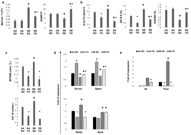
Sickle cell disease (SCD) is a genetic disorder of hemoglobin, leading to chronic hemolytic anemia and multiple organ damage. Among chronic organ complications, sickle cell bone disease (SBD) has a very high prevalence, resulting in long-term disability, chronic pain and fractures. Here, we evaluated the effects of ω-3 (fish oil-based, FD)-enriched diet vs. ω-6 (soybean oil-based, SD)- supplementation on murine SBD. We exposed SCD mice to recurrent hypoxia/reoxygenation (rec H/R), a consolidated model for SBD. In rec H/R SS mice, FD improves osteoblastogenesis/osteogenic activity by downregulating osteoclast activity via miR205 down-modulation and reduces both systemic and local inflammation. We also evaluated adipogenesis in both AA and SS mice fed with either SD or FD and exposed to rec H/R. FD reduced and reprogramed adipogenesis from white to brown adipocyte tissue (BAT) in bone compartments. This was supported by increased expression of uncoupling protein 1(UCP1), a BAT marker, and up-regulation of miR455, which promotes browning of white adipose tissue. Our findings provide new insights on the mechanism of action of ω-3 fatty acid supplementation on the pathogenesis of SBD and strengthen the rationale for ω-3 fatty acid dietary supplementation in SCD as a complementary therapeutic intervention.
Keywords: adipogenesis; bone histomorphometry; miRNAs; osteogenesis; sickle cell disease.
Conflict of interest statement
The authors declare no conflict of interest.
Figures


Similar articles
-
Hypoxia-reperfusion affects osteogenic lineage and promotes sickle cell bone disease.Blood. 2015 Nov 12;126(20):2320-8. doi: 10.1182/blood-2015-04-641969. Epub 2015 Sep 1. Blood. 2015. PMID: 26330244
-
Dietary ω-3 fatty acids protect against vasculopathy in a transgenic mouse model of sickle cell disease.Haematologica. 2015 Jul;100(7):870-80. doi: 10.3324/haematol.2015.124586. Epub 2015 May 1. Haematologica. 2015. PMID: 25934765 Free PMC article.
-
Maternal n-3 PUFA supplementation promotes fetal brown adipose tissue development through epigenetic modifications in C57BL/6 mice.Biochim Biophys Acta Mol Cell Biol Lipids. 2018 Dec;1863(12):1488-1497. doi: 10.1016/j.bbalip.2018.09.008. Epub 2018 Sep 25. Biochim Biophys Acta Mol Cell Biol Lipids. 2018. PMID: 30266429 Free PMC article.
-
Biochemical and therapeutic effects of Omega-3 fatty acids in sickle cell disease.Complement Ther Med. 2020 Aug;52:102482. doi: 10.1016/j.ctim.2020.102482. Epub 2020 Jun 9. Complement Ther Med. 2020. PMID: 32951732 Review.
-
Mechanisms of Bone Impairment in Sickle Bone Disease.Int J Environ Res Public Health. 2021 Feb 13;18(4):1832. doi: 10.3390/ijerph18041832. Int J Environ Res Public Health. 2021. PMID: 33668588 Free PMC article. Review.
Cited by
-
Crosstalk between Lipid Metabolism and Bone Homeostasis: Exploring Intricate Signaling Relationships.Research (Wash D C). 2024 Aug 20;7:0447. doi: 10.34133/research.0447. eCollection 2024. Research (Wash D C). 2024. PMID: 39165638 Free PMC article.
-
Targeting sickle cell pathobiology and pain with novel transdermal curcumin.PNAS Nexus. 2025 Feb 13;4(2):pgaf053. doi: 10.1093/pnasnexus/pgaf053. eCollection 2025 Feb. PNAS Nexus. 2025. PMID: 40007577 Free PMC article.
-
Transfusional Approach in Multi-Ethnic Sickle Cell Patients: Real-World Practice Data From a Multicenter Survey in Italy.Front Med (Lausanne). 2022 Mar 16;9:832154. doi: 10.3389/fmed.2022.832154. eCollection 2022. Front Med (Lausanne). 2022. PMID: 35372393 Free PMC article.
-
Feasibility trial for the management of severe acute malnutrition in older children with sickle cell anemia in Nigeria.Blood Adv. 2023 Oct 24;7(20):6024-6034. doi: 10.1182/bloodadvances.2023010789. Blood Adv. 2023. PMID: 37428866 Free PMC article. Clinical Trial.
-
Bone Disease among Children with Sickle Cell Disease: A Scoping Review of Incidence and Interventions.J Multidiscip Healthc. 2024 Jul 9;17:3235-3246. doi: 10.2147/JMDH.S475371. eCollection 2024. J Multidiscip Healthc. 2024. PMID: 39006879 Free PMC article.
References
-
- Maitra P., Caughey M., Robinson L., Desai P.C., Jones S., Nouraie M., Gladwin M.T., Hinderliter A., Cai J., Ataga K.I. Risk factors for mortality in adult patients with sickle cell disease: A meta-analysis of studies in North America and Europe. Haematology. 2017;102:626–636. doi: 10.3324/haematol.2016.153791. - DOI - PMC - PubMed
-
- Murray C.J.L., Vos T., Lozano R., Naghavi M., Flaxman A.D., Michaud C., Ezzati M., Shibuya K., Salomon J.A., Abdalla S., et al. Disability-adjusted life years (DALYs) for 291 diseases and injuries in 21 regions, 1990–2010: A systematic analysis for the Global Burden of Disease Study 2010. Lancet. 2012;380:2197–2223. doi: 10.1016/S0140-6736(12)61689-4. - DOI - PubMed
LinkOut - more resources
Full Text Sources
Research Materials
