Review
. 2021 May 18;26(10):3007.
doi: 10.3390/molecules26103007.

Bone Grafts and Substitutes in Dentistry: A Review of Current Trends and Developments

Affiliations
Review

Bone Grafts and Substitutes in Dentistry: A Review of Current Trends and Developments

Rusin Zhao et al. Molecules. .

Abstract

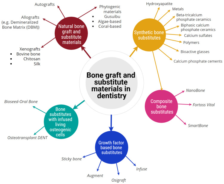
After tooth loss, bone resorption is irreversible, leaving the area without adequate bone volume for successful implant treatment. Bone grafting is the only solution to reverse dental bone loss and is a well-accepted procedure required in one in every four dental implants. Research and development in materials, design and fabrication technologies have expanded over the years to achieve successful and long-lasting dental implants for tooth substitution. This review will critically present the various dental bone graft and substitute materials that have been used to achieve a successful dental implant. The article also reviews the properties of dental bone grafts and various dental bone substitutes that have been studied or are currently available commercially. The various classifications of bone grafts and substitutes, including natural and synthetic materials, are critically presented, and available commercial products in each category are discussed. Different bone substitute materials, including metals, ceramics, polymers, or their combinations, and their chemical, physical, and biocompatibility properties are explored. Limitations of the available materials are presented, and areas which require further research and development are highlighted. Tissue engineering hybrid constructions with enhanced bone regeneration ability, such as cell-based or growth factor-based bone substitutes, are discussed as an emerging area of development.

Keywords: bone defects; bone graft; bone reconstruction; bone tissue engineering; dental implant; natural and synthetic bone substitutes; replacing tooth loss.


Conflict of interest statement

The authors declare no conflict of interest.

Figures

Figure 1
Figure 1
Use of structural scaffolds to restore bony defects. Diagram shows placement of a bone graft scaffold within a bony defect in alveolar bone following surgical generation of an access flap.
Figure 2
Figure 2
Classification of bone graft and substitute materials used in dentistry, broadly classified into five categories and showing their associated sub-categories.
Figure 3
Figure 3
Clinical images illustrating the pre- and post-operative (post-op) procedures of an edentulous patient treated with an implant with guided bone tissue regeneration. Images from left to right show: (a) the edentulous site after the tooth was extracted; bone defect in the form of buccal concavity is visible in the apical aspect of 24 (yellow arrow). (b) Occlusal shot of the edentulous site showing the buccal concavity. (c) The correct positioning of the implant (Straumann Bone Level Tapered 3.3 × 10 mm implant). (d) Proper bucco-palatal positioning of the implant. (e) Decorticated area to prior to placement of the bone and complete absence of the buccal bone at the apical part of the implant. (f) The placement of Straumann Flex Membrane fixed and stabilized by tacks (AutoTac by BioHorizons Canada). (g) Applying bone graft particles comprising of a mixture of Allograft and Xenograft (both from Straumann®) packed at the buccal bone defect. (h) Periosteal sutures used to stabilize and fix the bone graft inside the membrane, which ensures immobilization of graft resulting in optimal bone regeneration vs. fibrous tissue formation. (i) Primary closure of the site. (j) Showing post-op. Primary closure is intact. (k) The implant after second stage and osseointegration check. (l) Five months later, post-op cone beam computed tomography (CBCT) illustrating the final bone healing prior to second stage and osseointegration check of the implant. The post-op CBCT revealing a gain of over 5 mm of bone (courtesy of Dr. Mohammad A. Javaid, Periodontist, British Columbia, Canada).
Figure 4
Figure 4
(A) Use of a titanium mesh as a structural scaffold and physical barrier in GBR for prevention of soft tissue cell migration and promotion of bone regeneration. (B) Use of a barrier membrane in GBR.

References

    1. Cypher T.J., Grossman J.P. Biological principles of bone graft healing. J. Foot Ankle Surg. 1996;35:413–417. doi: 10.1016/S1067-2516(96)80061-5. - DOI - PubMed
    1. Elsalanty M.E., Genecov D.G. Bone Grafts in Craniofacial Surgery. Craniomaxillofacial Trauma Reconstr. 2009;2:125–134. doi: 10.1055/s-0029-1215875. - DOI - PMC - PubMed
    1. Fernandez de Grado G., Keller L., Idoux-Gillet Y., Wagner Q., Musset A.M., Benkirane-Jessel N., Bornert F., Offner D. Bone substitutes: A review of their characteristics, clinical use, and perspectives for large bone defects management. J. Tissue Eng. 2018;9:2041731418776819. doi: 10.1177/2041731418776819. - DOI - PMC - PubMed
    1. Abhay S., Haines S.J. Repairing holes in the head: A history of cranioplasty. Neurosurgery. 1997;40:588–603. - PubMed
    1. Kumar P., Fathima G., Vinitha B. Bone grafts in dentistry. J. Pharm. Bioallied Sci. 2013;5:125–127. doi: 10.4103/0975-7406.113312. - DOI - PMC - PubMed

MeSH terms

Substances