Review
. 2021 May 18;13(5):936.
doi: 10.3390/v13050936.

Overview of Bat and Wildlife Coronavirus Surveillance in Africa: A Framework for Global Investigations

Affiliations
Review

Overview of Bat and Wildlife Coronavirus Surveillance in Africa: A Framework for Global Investigations

Marike Geldenhuys et al. Viruses. .

Abstract

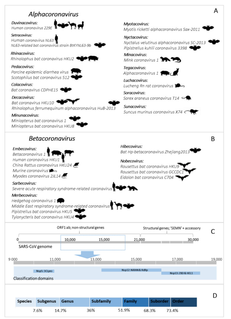
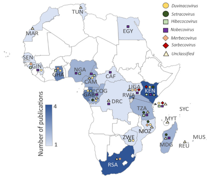
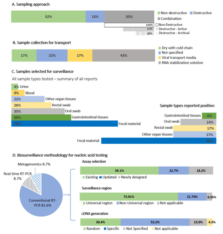
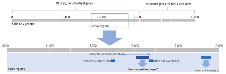
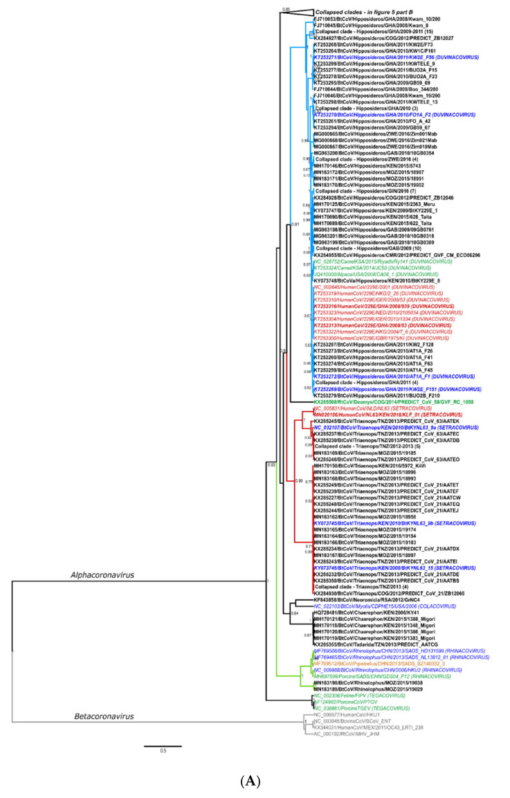
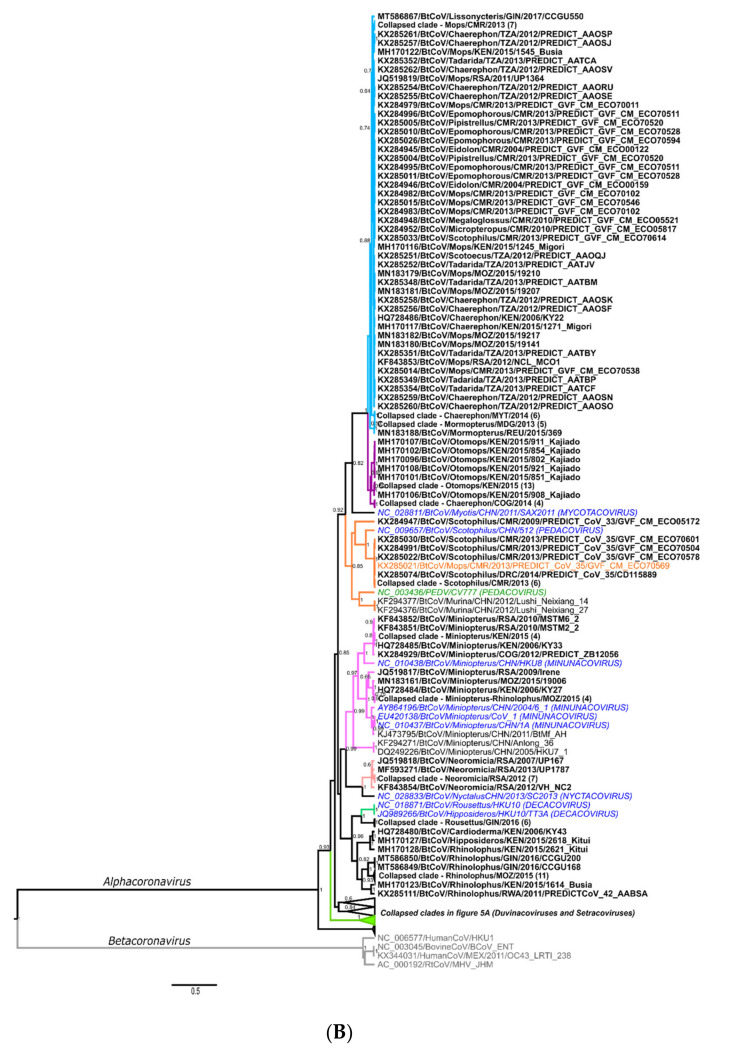
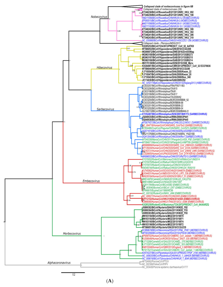
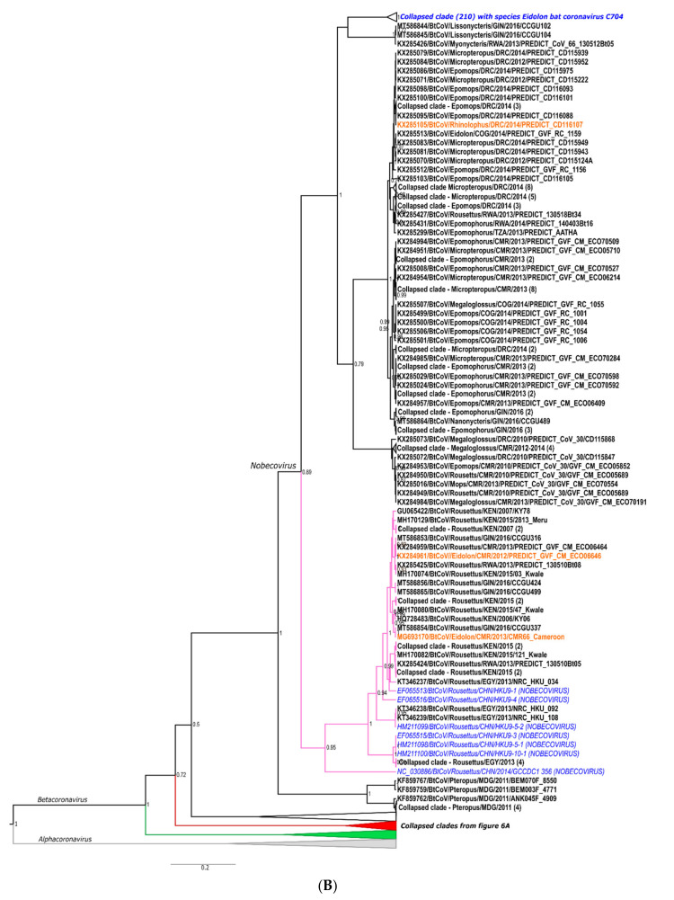
The ongoing coronavirus disease 2019 (COVID-19) pandemic has had devastating health and socio-economic impacts. Human activities, especially at the wildlife interphase, are at the core of forces driving the emergence of new viral agents. Global surveillance activities have identified bats as the natural hosts of diverse coronaviruses, with other domestic and wildlife animal species possibly acting as intermediate or spillover hosts. The African continent is confronted by several factors that challenge prevention and response to novel disease emergences, such as high species diversity, inadequate health systems, and drastic social and ecosystem changes. We reviewed published animal coronavirus surveillance studies conducted in Africa, specifically summarizing surveillance approaches, species numbers tested, and findings. Far more surveillance has been initiated among bat populations than other wildlife and domestic animals, with nearly 26,000 bat individuals tested. Though coronaviruses have been identified from approximately 7% of the total bats tested, surveillance among other animals identified coronaviruses in less than 1%. In addition to a large undescribed diversity, sequences related to four of the seven human coronaviruses have been reported from African bats. The review highlights research gaps and the disparity in surveillance efforts between different animal groups (particularly potential spillover hosts) and concludes with proposed strategies for improved future biosurveillance.

Keywords: Africa; African bat coronaviruses; COVID-19; HCoV-229E; HCoV-NL63; MERS-CoV; SARS-CoV; SARS-CoV 2; bat; biosurveillance; coronaviruses; domestic animals; emerging; surveillance; surveillance strategies; wildlife.


Conflict of interest statement

The authors declare no conflict of interest. The funders had no role in the design of the study; in the collection, analyses, or interpretation of data; in the writing of the manuscript, or in the decision to publish the results.

Figures

Figure 1
Figure 1
(A,B) Current coronavirus subgenera (bold) and species of the Alphacoronavirus and Betacoronavirus genera. The images indicate host species associated with the virus species. Figure constructed with the species listed on the 2019 Release of the ICTV Virus Taxonomy 9th Report MSL#35: (Available at https://talk.ictvonline.org/ictv-reports/ictv_9th_report/positive-sense-rna-viruses-2011/w/posrna_viruses/222/coronaviridae accessed on 12 December 2020). (C) Representation of the coronavirus genome (based on the reference genome NC_004718.3 SARS coronavirus Tor2) depicting the locations of important domains for classification of species (NSP5 (3CLpro), NSP12 (NiRAN and RdRp), and NSP13 (ZBD and HEL1)). (D) Thresholds of the taxonomic demarcation criteria [24]. Novel viruses are part of a taxonomic level if the divergence within the five concatenated replicase domains is less than the indicated amino acid percentage.
Figure 2
Figure 2
Published bat coronavirus surveillance studies per country (shading denoting the number of publications). Symbols in the key above the map represent different coronaviruses detected in the respective countries: Duvinacovirus as a yellow circle (HCoV229E-related viruses), Setracoronavirus as a dark green circle (HCoVNL63-related viruses), Sarbecoviruses as a red diamond (HCoV-SARS-related viruses), Merbecoviruses as an orange diamond (HCoV-MERS-related viruses), Nobecoviruses as a purple square, Hibecoviruses as a green square, and unclassified viruses as a black triangle. Further details on coronaviruses identified can be reviewed in Table S4. Three-letter ISO country code abbreviations are shown on the map.
Figure 3
Figure 3
A summary of coronavirus sampling approaches and methodology. (A) The sampling approaches of the 23 primary surveillance reports. Combination studies are split into those employing new or archival destructive sampling. (B) Sample preservation methods. (C) Sample types selected for surveillance and samples testing positive. (D) Biosurveillance methodology for nucleic acid testing, percentage of studies using conventional, real-time, or metagenomic approaches. The conventional assays were further split into existing assays from the literature, updated exiting assays, or whether new assays were developed. The percentages of studies targeting the ‘universal surveillance region’ (see text for an explanation) contrast to those using different genome regions, and whether specific or random primers were chosen for cDNA preparation.
Figure 4
Figure 4
Representation of the coronavirus genome (based on the reference genome NC_004718.3 SARS coronavirus Tor2) depicting the assay regions. The assays corresponding to this universal region included in Tong et al. [26], de Souza Luna [62], Geldenhuys et al. [42] and Geldenhuys et al. [32] (based on primers from Woo et al. [63]), Razanajatovo et al. [47] (based on Poon et al. [14]), Shehata et al. [27], Waruhiu et al. [29] (based on Watanabe et al. [64]), Chu et al. [65], Gouilh et al., [33]. The RdRp grouping units (RGU) amplification region by Drexler et al. [66] is indicated with the line and arrows.
Figure 5
Figure 5
(A,B): Alphacoronavirus Bayesian phylogeny of the RdRp partial region (corresponding to approximately 15,200–15,400 nt of the coronavirus genome). Clades collapsed in A are shown in B (and vice versa). To include the maximum number of sequences, sequence lengths were trimmed to a generally useable length of 260 nucleotides. Sequences resulting in shorter lengths were omitted. Sequences in italics indicate formally recognized species (subgenera indicated in capital letters at the end of sequence names); sequences in bold originate in Africa; red highlights human viruses; green indicate non-bat animal hosts; blue/italics indicate formally recognized bat species; orange indicate viral detections from hosts not typically associated with a particular group of coronaviruses. All sequence names were edited to conform to the correct convention, with the modification of the unique sequence identifier listed last due to convenience. Only posterior probabilities of greater than 0.5 are indicated. No unpublished sequences are shown.
Figure 5
Figure 5
(A,B): Alphacoronavirus Bayesian phylogeny of the RdRp partial region (corresponding to approximately 15,200–15,400 nt of the coronavirus genome). Clades collapsed in A are shown in B (and vice versa). To include the maximum number of sequences, sequence lengths were trimmed to a generally useable length of 260 nucleotides. Sequences resulting in shorter lengths were omitted. Sequences in italics indicate formally recognized species (subgenera indicated in capital letters at the end of sequence names); sequences in bold originate in Africa; red highlights human viruses; green indicate non-bat animal hosts; blue/italics indicate formally recognized bat species; orange indicate viral detections from hosts not typically associated with a particular group of coronaviruses. All sequence names were edited to conform to the correct convention, with the modification of the unique sequence identifier listed last due to convenience. Only posterior probabilities of greater than 0.5 are indicated. No unpublished sequences are shown.
Figure 6
Figure 6
(A,B): Bayesian Betacoronavirus phylogeny of a 294-nucleotide sequence region of the RdRp gene. Shorter sequences were omitted. Clades collapsed in A are shown in B (and vice versa), and the collapsed clade of Eidolon nobecoviruses may be viewed in Figure S2). Sequences in italics indicate formally recognized species (subgenera are indicated in capital letters at the end of sequence names); sequences in bold originate in Africa; red highlights human viruses; green indicate non-bat animal hosts; blue/italics indicate formally recognized bat species; orange indicate viral detections from hosts not typically associated with a particular group of coronaviruses. All sequence names were edited to conform to the correct convention, with the modification of the unique sequence identifier listed last due to convenience. Only posterior probabilities of greater than 0.5 are indicated. No unpublished sequences were included.
Figure 6
Figure 6
(A,B): Bayesian Betacoronavirus phylogeny of a 294-nucleotide sequence region of the RdRp gene. Shorter sequences were omitted. Clades collapsed in A are shown in B (and vice versa), and the collapsed clade of Eidolon nobecoviruses may be viewed in Figure S2). Sequences in italics indicate formally recognized species (subgenera are indicated in capital letters at the end of sequence names); sequences in bold originate in Africa; red highlights human viruses; green indicate non-bat animal hosts; blue/italics indicate formally recognized bat species; orange indicate viral detections from hosts not typically associated with a particular group of coronaviruses. All sequence names were edited to conform to the correct convention, with the modification of the unique sequence identifier listed last due to convenience. Only posterior probabilities of greater than 0.5 are indicated. No unpublished sequences were included.

Similar articles

Cited by

References

    1. Ksiazek T.G., Erdman D., Goldsmith C.S., Zaki S.R., Peret T., Emery S., Tong S., Urbani C., Comer J.A., Lim W., et al. A novel coronavirus associated with severe acute respiratory syndrome. N. Engl. J. Med. 2003;348:1953–1966. doi: 10.1056/NEJMoa030781. - DOI - PubMed
    1. Berry M., Gamieldien J., Fielding B.C. Identification of new respiratory viruses in the new millennium. Viruses. 2015;7:996–1019. doi: 10.3390/v7030996. - DOI - PMC - PubMed
    1. Zhou P., Fan H., Lan T., Yang X.L., Shi W.F., Zhang W., Zhu Y., Zhang Y.W., Xie Q.M., Mani S., et al. Fatal swine acute diarrhoea syndrome caused by an HKU2-related coronavirus of bat origin. Nature. 2018;556:255–259. doi: 10.1038/s41586-018-0010-9. - DOI - PMC - PubMed
    1. Zhou L., Li Q.N., Su J.N., Chen G.H., Wu Z.X., Luo Y., Wu R.T., Sun Y., Lan T., Ma J.Y. The re-emerging of SADS-CoV infection in pig herds in Southern China. Transbound. Emerg. Dis. 2019;66:2180–2183. doi: 10.1111/tbed.13270. - DOI - PMC - PubMed
    1. Zhou P., Yang X.L., Wang X.G., Hu B., Zhang L., Zhang W., Si H.R., Zhu Y., Li B., Huang C.L., et al. A pneumonia outbreak associated with a new coronavirus of probable bat origin. Nature. 2020;579:270–273. doi: 10.1038/s41586-020-2012-7. - DOI - PMC - PubMed

Publication types

Substances

LinkOut - more resources