Case Reports
. 2021 May 18;22(10):5324.
doi: 10.3390/ijms22105324.

The Role of ZEB2 in Human CD8 T Lymphocytes: Clinical and Cellular Immune Profiling in Mowat-Wilson Syndrome

Affiliations
Case Reports

The Role of ZEB2 in Human CD8 T Lymphocytes: Clinical and Cellular Immune Profiling in Mowat-Wilson Syndrome

Katie Frith et al. Int J Mol Sci. .

Abstract

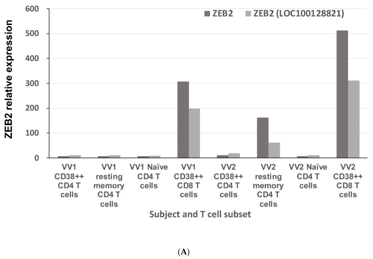
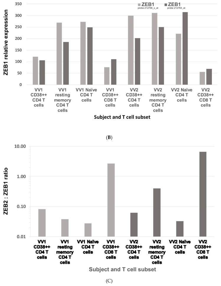
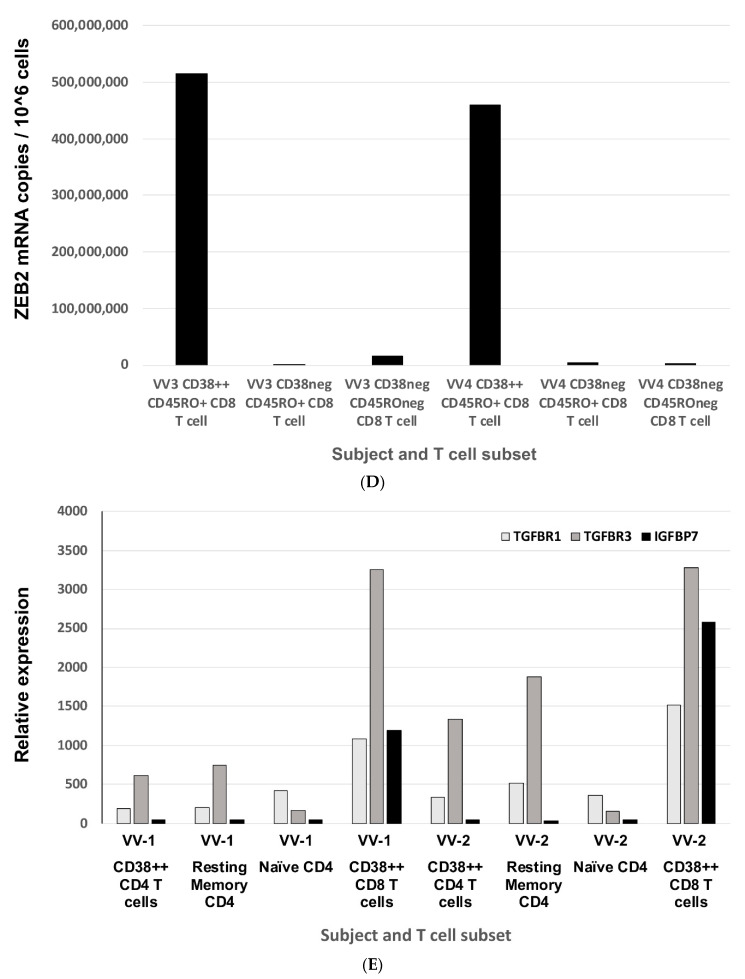
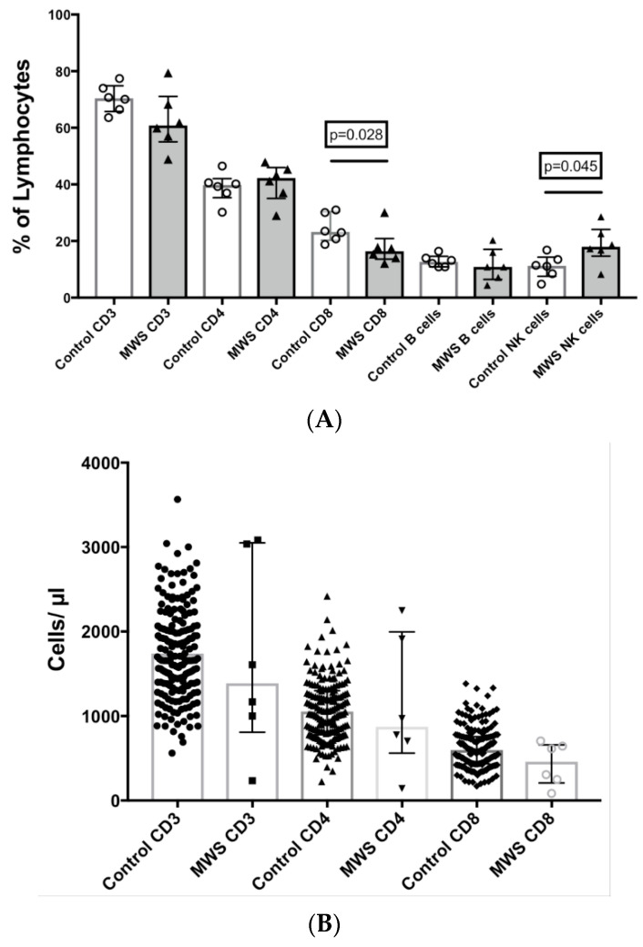
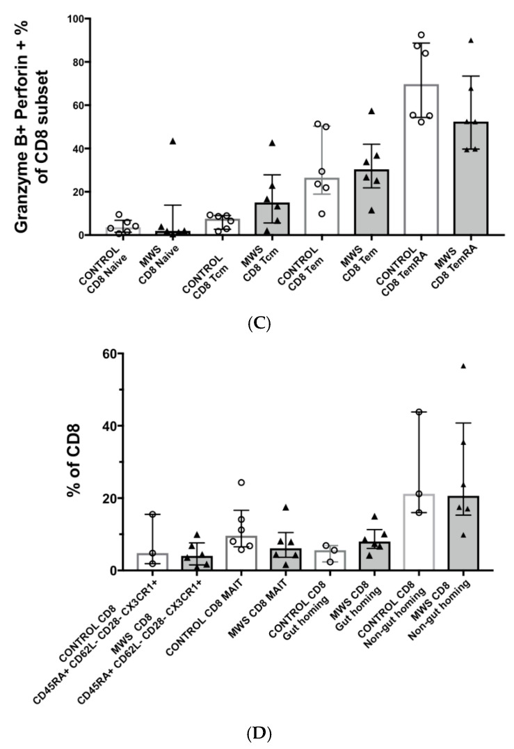
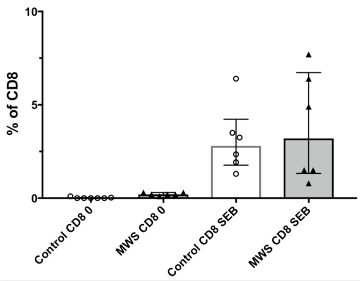
The Zeb2 gene encodes a transcription factor (ZEB2) that acts as an important immune mediator in mice, where it is expressed in early-activated effector CD8 T cells, and limits effector differentiation. Zeb2 homozygous knockout mice have deficits in CD8 T cells and NK cells. Mowat-Wilson syndrome (MWS) is a rare genetic disease resulting from heterozygous mutations in ZEB2 causing disease by haploinsufficiency. Whether ZEB2 exhibits similar expression patterns in human CD8 T cells is unknown, and MWS patients have not been comprehensively studied to identify changes in CD8 lymphocytes and NK cells, or manifestations of immunodeficiency. By using transcriptomic assessment, we demonstrated that ZEB2 is expressed in early-activated effector CD8 T cells of healthy human volunteers following vaccinia inoculation and found evidence of a role for TGFß-1/SMAD signaling in these cells. A broad immunological assessment of six genetically diagnosed MWS patients identified two patients with a history of recurrent sinopulmonary infections, one of whom had recurrent oral candidiasis, one with lymphopenia, two with thrombocytopenia and three with detectable anti-nuclear antibodies. Immunoglobulin levels, including functional antibody responses to protein and polysaccharide vaccination, were normal. The MWS patients had a significantly lower CD8 T cell subset as % of lymphocytes, compared to healthy controls (median 16.4% vs. 25%, p = 0.0048), and resulting increased CD4:CD8 ratio (2.6 vs. 1.8; p = 0.038). CD8 T cells responded normally to mitogen stimulation in vitro and memory CD8 T cells exhibited normal proportions of subsets with important tissue-specific homing markers and cytotoxic effector molecules. There was a trend towards a decrease in the CD8 T effector memory subset (3.3% vs. 5.9%; p = 0.19). NK cell subsets were normal. This is the first evidence that ZEB2 is expressed in early-activated human effector CD8 T cells, and that haploinsufficiency of ZEB2 in MWS patients had a slight effect on immune function, skewing T cells away from CD8 differentiation. To date there is insufficient evidence to support an immunodeficiency occurring in MWS patients.

Keywords: CD8 T cells; Mowat–Wilson syndrome; zinc finger E-box binding homeobox 2 gene (ZEB2).


Conflict of interest statement

The authors declare no conflict of interest.

Figures

Figure 1
Figure 1
Gene expression profiling of ZEB2 and ZEB1 RNA and TGF-ß1 signalling molecules in microarrays of early-activated effector CD38+++ CD8 T cells following vaccinia inoculation. Four purified T cell subsets were cell sorted from fresh PBMC: CD38++ CD4 T cells; resting memory CD4 T cells; naïve CD4 T cells and CD38++ CD8 T cells from each of two healthy adult donors, VV1 and VV2. RNA levels for each subset were normalised and calculated in Affymetrix MAS 5.0 software. (A) Elevated expression of ZEB2 in microarrays of early-activated human CD38++ CD8 effector T cells, 2-weeks post-inoculation of vaccinia virus. The light grey bars are for probe set 235593_at, annotated as specific for Zeb2 (Accession No. AL546529) and the dark grey bars are for probe set 203603_s_at (Accession No. NM_014795), also annotated as Zeb2. (B) Reduced expression of ZEB1 in microarrays of early-activated human CD38++ CD8 effector T cells, 2-weeks post-inoculation of vaccinia virus. The light grey bars are for probe set 212758_s_at, annotated as specific for Zeb1 (Accession No. AI373166) and the dark grey bars are for probe set 212764_at (Accession No. AI806174), also annotated as Zeb1. (C) Elevated ZEB2 and lower ZEB1 expression in early-activated human CD38++ CD8 antiviral effector cells leads to a ratio of >1 in those cells, compared to other T cells. (D) Elevated levels of ZEB2 mRNA were confirmed by quantitative real-time RT-PCR of purified early-activated human CD38++ CD8 effector T cells, from cryopreserved PBMC from two different donors, 2-weeks post-inoculation of vaccinia virus. (E) Elevated expression of TGF-ß1 receptors (TGFBR1 and TGFBR3) and IGFBP7 in early-activated human CD38++ CD8 antiviral effector cells, by microarray analysis.
Figure 1
Figure 1
Gene expression profiling of ZEB2 and ZEB1 RNA and TGF-ß1 signalling molecules in microarrays of early-activated effector CD38+++ CD8 T cells following vaccinia inoculation. Four purified T cell subsets were cell sorted from fresh PBMC: CD38++ CD4 T cells; resting memory CD4 T cells; naïve CD4 T cells and CD38++ CD8 T cells from each of two healthy adult donors, VV1 and VV2. RNA levels for each subset were normalised and calculated in Affymetrix MAS 5.0 software. (A) Elevated expression of ZEB2 in microarrays of early-activated human CD38++ CD8 effector T cells, 2-weeks post-inoculation of vaccinia virus. The light grey bars are for probe set 235593_at, annotated as specific for Zeb2 (Accession No. AL546529) and the dark grey bars are for probe set 203603_s_at (Accession No. NM_014795), also annotated as Zeb2. (B) Reduced expression of ZEB1 in microarrays of early-activated human CD38++ CD8 effector T cells, 2-weeks post-inoculation of vaccinia virus. The light grey bars are for probe set 212758_s_at, annotated as specific for Zeb1 (Accession No. AI373166) and the dark grey bars are for probe set 212764_at (Accession No. AI806174), also annotated as Zeb1. (C) Elevated ZEB2 and lower ZEB1 expression in early-activated human CD38++ CD8 antiviral effector cells leads to a ratio of >1 in those cells, compared to other T cells. (D) Elevated levels of ZEB2 mRNA were confirmed by quantitative real-time RT-PCR of purified early-activated human CD38++ CD8 effector T cells, from cryopreserved PBMC from two different donors, 2-weeks post-inoculation of vaccinia virus. (E) Elevated expression of TGF-ß1 receptors (TGFBR1 and TGFBR3) and IGFBP7 in early-activated human CD38++ CD8 antiviral effector cells, by microarray analysis.
Figure 2
Figure 2
Comparison of lymphocyte subsets in peripheral blood between controls and patients with Mowat–Wilson syndrome. (A) T cell (CD3+, CD4+ and CD8+), B cell (CD19+) and NK cell (CD56+) subsets as % of total lymphocytes. (B) Comparison of T cell counts (cells/μL) between controls and patients with Mowat–Wilson syndrome.
Figure 3
Figure 3
Comparison of CD8 T cell subsets in peripheral blood between controls and patients with Mowat–Wilson syndrome. (A) Comparison of naïve (CD45RO−CD62L+), central memory (CD45RO+CD62L+), effector memory (CD45RO+CD62L−) and TEMRA (CD45RO−CD62L−) subsets of CD8 T cells between controls and patients with Mowat–Wilson syndrome. Subsets are expressed as % of total CD8 T cells. (B) Comparison of CD8 T effector memory (CD45RO+CD62L−) subsets between historical controls, same day controls and patients with Mowat–Wilson syndrome. Subsets are expressed as % of total lymphocytes. (C) Comparison of cytotoxic CD8 T effector cells as % of naïve (CD45RO−CD62L+), central memory (CD45RO+CD62L+), effector memory (CD45RO+CD62L−) and TEMRA (CD45RO−CD62L−) subsets, respectively, controls and patients with Mowat–Wilson syndrome. Granzyme B+ Perforin+ cells are expressed as % of each subset of CD8+ T lymphocytes. (D) Comparison of terminally differentiated (CD45RA+CD62L−CD28-CX3CR1+) CD8 T cells, MAIT (CD45RO+CD161highCCR6+CD127high) cells and gut-homing (CD45RO+CD49d+Integrinß7+) CD8 T cells between controls and patients with Mowat–Wilson syndrome. Subsets are expressed as % of total CD8 T cells.
Figure 3
Figure 3
Comparison of CD8 T cell subsets in peripheral blood between controls and patients with Mowat–Wilson syndrome. (A) Comparison of naïve (CD45RO−CD62L+), central memory (CD45RO+CD62L+), effector memory (CD45RO+CD62L−) and TEMRA (CD45RO−CD62L−) subsets of CD8 T cells between controls and patients with Mowat–Wilson syndrome. Subsets are expressed as % of total CD8 T cells. (B) Comparison of CD8 T effector memory (CD45RO+CD62L−) subsets between historical controls, same day controls and patients with Mowat–Wilson syndrome. Subsets are expressed as % of total lymphocytes. (C) Comparison of cytotoxic CD8 T effector cells as % of naïve (CD45RO−CD62L+), central memory (CD45RO+CD62L+), effector memory (CD45RO+CD62L−) and TEMRA (CD45RO−CD62L−) subsets, respectively, controls and patients with Mowat–Wilson syndrome. Granzyme B+ Perforin+ cells are expressed as % of each subset of CD8+ T lymphocytes. (D) Comparison of terminally differentiated (CD45RA+CD62L−CD28-CX3CR1+) CD8 T cells, MAIT (CD45RO+CD161highCCR6+CD127high) cells and gut-homing (CD45RO+CD49d+Integrinß7+) CD8 T cells between controls and patients with Mowat–Wilson syndrome. Subsets are expressed as % of total CD8 T cells.
Figure 4
Figure 4
Lymphocyte function of CD8 T cells in response to the polyclonal mitogen SEB. Cells were incubated for 2 days with or without SEB, and the % of CD8 T cells that responded by upregulated coexpression of CD25 and CD134.

Similar articles

Cited by

References

    1. Mowat D.R., Croaker G.D., Cass D.T., Kerr B.A., Chaitow J., Ades L.C., Chia N.L., Wilson M.J. Hirschsprung disease, microcephaly, mental retardation, and characteristic facial features: Delineation of a new syndrome and identification of a locus at chromosome 2q22-q23. J. Med. Genet. 1998;35:617–623. doi: 10.1136/jmg.35.8.617. - DOI - PMC - PubMed
    1. Ivanovski I., Djuric O., Caraffi S.G., Santodirocco D., Pollazzon M., Rosato S., Cordelli D.M., Abdalla E., Accorsi P., Adam M.P., et al. Phenotype and genotype of 87 patients with Mowat–Wilson syndrome and recommendations for care. Genet. Med. 2018;20:965–975. doi: 10.1038/gim.2017.221. - DOI - PubMed
    1. Ho S., Luk H.-M., Chung B.H.-Y., Fung J.L.-F., Mak H.H.-Y., Lo I.F.M. Mowat–Wilson syndrome in a Chinese population: A case series. Am. J. Med. Genet. Part A. 2020;182:1336–1341. doi: 10.1002/ajmg.a.61557. - DOI - PubMed
    1. Zweier C., Thiel C.T., Dufke A., Crow Y.J., Meinecke P., Suri M., Ala-Mello S., Beemer F., Bernasconi S., Bianchi P., et al. Clinical and Mutational Spectrum of Mowat–Wilson Syndrome. Eur. J. Med. Genet. 2005;48:97–111. doi: 10.1016/j.ejmg.2005.01.003. - DOI - PubMed
    1. Omilusik K.D., Best J.A., Yu B., Goossens S., Weidemann A., Nguyen J.V., Seuntjens E., Stryjewska A., Zweier C., Roychoudhuri R., et al. Transcriptional repressor ZEB2 promotes terminal differentiation of CD8+ effector and memory T cell populations during infection. J. Exp. Med. 2015;212:2027–2039. doi: 10.1084/jem.20150194. - DOI - PMC - PubMed

Publication types

MeSH terms

Substances

Supplementary concepts