. 2021 May 25;22(11):5571.
doi: 10.3390/ijms22115571.

Adenosine and Cordycepin Accelerate Tissue Remodeling Process through Adenosine Receptor Mediated Wnt/β-Catenin Pathway Stimulation by Regulating GSK3b Activity

Affiliations

Adenosine and Cordycepin Accelerate Tissue Remodeling Process through Adenosine Receptor Mediated Wnt/β-Catenin Pathway Stimulation by Regulating GSK3b Activity

Jaeyoon Kim et al. Int J Mol Sci. .

Abstract

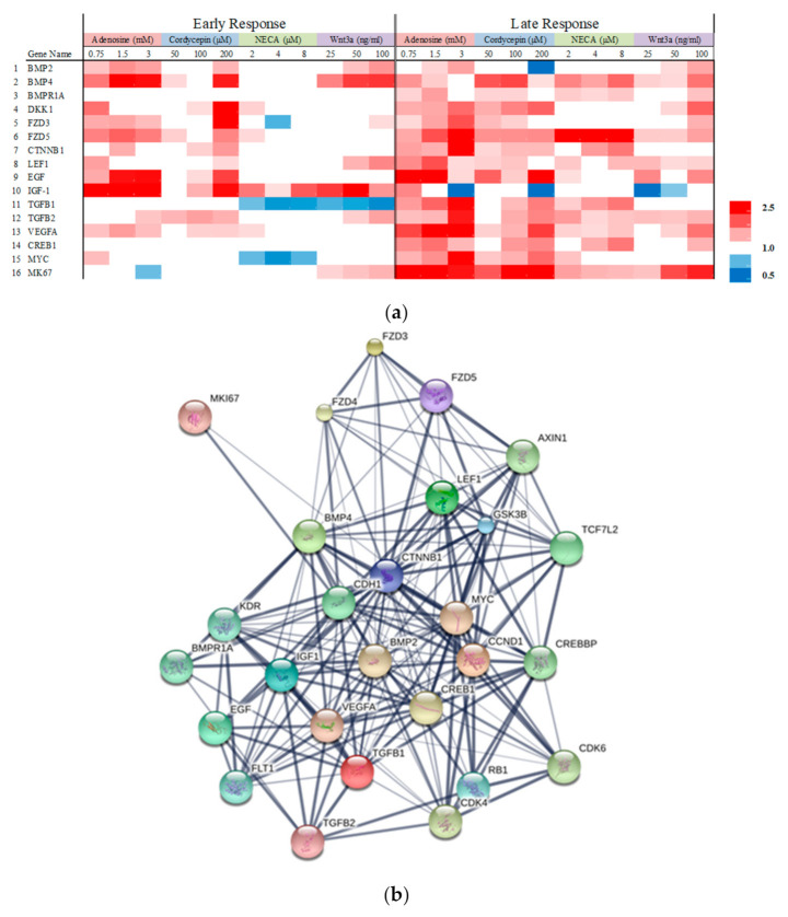
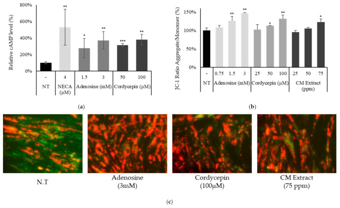
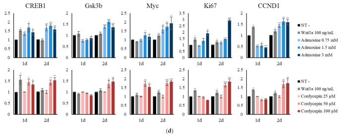
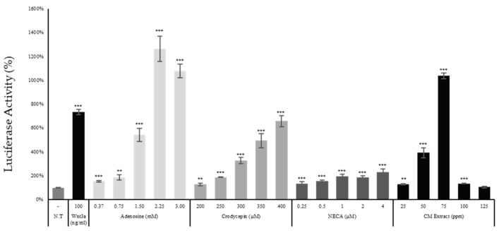
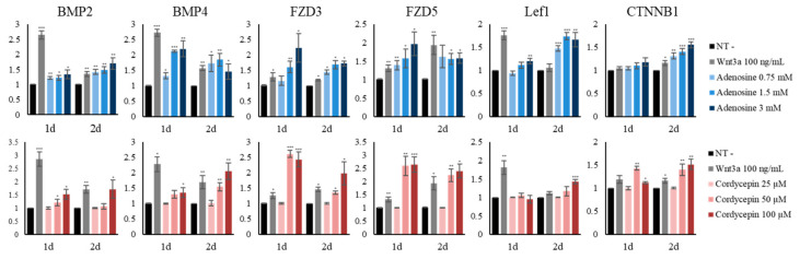
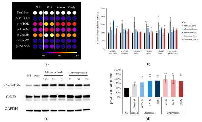
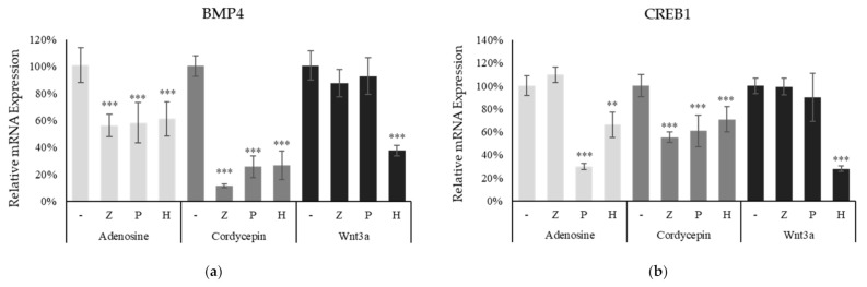
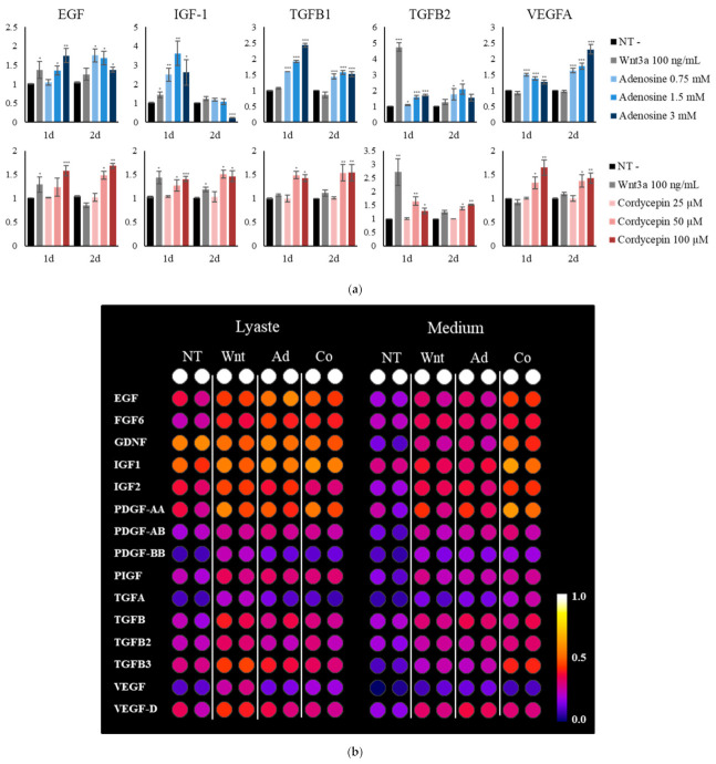
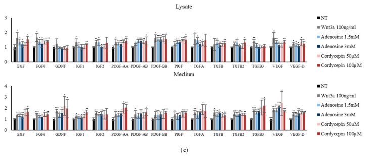
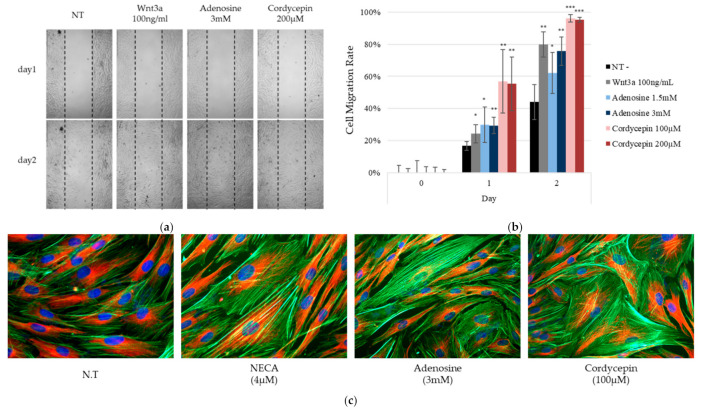
Adenosine is a cellular metabolite with diverse derivatives that possesses a wide range of physiological roles. We investigated the molecular mechanisms of adenosine and cordycepin for their promoting effects in wound-healing process. The mitochondrial energy metabolism and cell proliferation markers, cAMP responsive element binding protein 1 (CREB1) and Ki67, were enhanced by adenosine and cordycepin in cultured dermal fibroblasts. Adenosine and cordycepin stimulated adenosine receptor signaling via elevated cAMP. The phosphorylation of mitogen-activated protein kinase kinase (MEK) 1/2, mammalian target of rapamycin (mTOR) and glycogen synthase kinase 3 beta (Gsk3b) and Wnt target genes such as bone morphogenetic protein (BMP) 2/4 and lymphoid enhancer binding factor (Lef) 1 were activated. The enhanced gene expression by adenosine and cordycepin was abrogated by adenosine A2A and A2B receptor inhibitors, ZM241385 and PSH603, and protein kinase A (PKA) inhibitor H89, indicating the involvement of adenosine receptor A2A, A2B and PKA. As a result of Wnt/β-catenin pathway activation, the secretion of growth factors such as insulin-like growth factor (IGF)-1 and transforming growth factor beta (TGFβ) 3 was increased, previously reported to facilitate the wound healing process. In addition, in vitro fibroblast migration was also increased, demonstrating their possible roles in facilitating the wound healing process. In conclusion, our data strongly demonstrate that adenosine and cordycepin stimulate the Wnt/β-catenin signaling through the activation of adenosine receptor, possibly promoting the tissue remodeling process and suggest their therapeutic potential for treating skin wounds.

Keywords: Wnt/β-catenin pathway; adenosine; cordycepin; wound healing.


Conflict of interest statement

The authors declare no conflict of interest.

Figures

Figure 1
Figure 1
Chemical structures of adenosine, cordycepin, and NECA and the mRNA expression profile of adenosine receptor subtypes. (a) Chemical structures. (b) The expression of four adenosine receptor subtypes, A1 (ADORA1), A2A (ADORA2A), A2B (ADORA2B), and A3 (ADORA3) was examined using quantitative RT-PCR analysis in cultured human dermal fibroblast. Ct values for each receptor subtype were indicated.
Figure 2
Figure 2
The changes in mRNA expression profiles by adenosine analogues and recombinant wnt3a in cultured human dermal fibroblasts. Cells were treated with adenosine, cordycepin, NECA, and wnt3a for one day (early response) and two days (late response) and mRNA expression was examined by RT-PCR. (a) Sixteen genes with more than a two-fold increase and statistical significance (p < 0.05) was displayed, red and blue color mean increase and decrease in the mRNA expression, respectively, (b) further investigated by protein cluster analysis.
Figure 3
Figure 3
Adenosine and cordycepin activated adenosine receptor pathway in cultured human dermal fibroblast. Cells were treated with adenosine, cordycepin, NECA, and CM extract. (a) After 4 h treatment, intracellular cAMP levels were measured. NECA was used as a positive control for adenosine receptor activation. (b) Mitochondrial membrane potential was measured after 4 h treatment with adenosine, cordycepin, and CM extract. (c) Representative photos of JC-1 staining. JC-1 monomer form was seen as green and aggregate form as red by fluorescent microscopy. (d) The mRNA expression levels of CREB1, GSK3B, MYC, Ki67, and CCND1 were measured after treatment with adenosine and cordycepin for one day and two days. N.T, non-treated control; Significantly different compared with N.T (* p < 0.05, ** p < 0.01, *** p < 0.001).
Figure 3
Figure 3
Adenosine and cordycepin activated adenosine receptor pathway in cultured human dermal fibroblast. Cells were treated with adenosine, cordycepin, NECA, and CM extract. (a) After 4 h treatment, intracellular cAMP levels were measured. NECA was used as a positive control for adenosine receptor activation. (b) Mitochondrial membrane potential was measured after 4 h treatment with adenosine, cordycepin, and CM extract. (c) Representative photos of JC-1 staining. JC-1 monomer form was seen as green and aggregate form as red by fluorescent microscopy. (d) The mRNA expression levels of CREB1, GSK3B, MYC, Ki67, and CCND1 were measured after treatment with adenosine and cordycepin for one day and two days. N.T, non-treated control; Significantly different compared with N.T (* p < 0.05, ** p < 0.01, *** p < 0.001).
Figure 4
Figure 4
Adenosine, cordycepin, and NECA stimulated Wnt reporter activity. The Wnt reporter WRHEK293A cells were treated with adenosine, cordycepin, NECA, and CM extract and the luciferase activity was measured. N.T, non-treated control; significantly different compared with N.T (* p < 0.05, ** p < 0.01, *** p < 0.001).
Figure 5
Figure 5
Adenosine and cordycepin activated Wnt/β-catenin signaling pathway in cultured human fibroblasts. The expression levels of 6 Wnt/β-catenin pathway related genes (BMP2/4, FZD3/5, LEF1 and CTNNB1) were assessed in fibroblasts treated with adenosine and cordycepin for one day and two days. Recombinant wnt3a was used as a positive control. The data represent the means of six independent samples. N.T, non-treated control; significantly different compared with N.T (* p < 0.05, ** p < 0.01, *** p < 0.001).
Figure 6
Figure 6
Adenosine and cordycepin increased phosphorylation of Gsk3b on Ser9 in cultured human fibroblasts. (a,b) Cells were treated with adenosine and cordycepin for one day. Whole cell lysates were analyzed by immunoblotting to determine the phosphorylation levels of MAP kinases following manufacturer’s instruction. A total of 17 MAP kinases were analyzed. Six kinases with significantly increased phosphorylation were displayed. (c) The levels of phospho-Gsk3b, Gsk3b were further investigated by Western blotting after 4 h treatment with adenosine and cordycepin. (d) The ratio of p-Gsk3b/Gsk3b was calculated. The data represent the means of six independent samples. N.T, non-treated control; significantly different compared with N.T (* p < 0.05, ** p < 0.01, *** p < 0.001).
Figure 7
Figure 7
Activation of Wnt/β-catenin pathway by adenosine and cordycepin was abolished in the presence of adenosine receptor antagonists and PKA inhibitor. The cells were treated with adenosine and cordycepin in the presence of various inhibitors (ZM241385, PSB603 and H89) for 24 h, harvested, and the mRNA expression levels of BMP4 (a) and CREB1 (b) were measured by RT-PCR. The data represent the means of six independent samples. Significantly different compared with each single adenosine, cordycepin and wnt3a treatment. (** p < 0.01, *** p < 0.001); Adenosine (3 mM); Cordycepin (100 µM); Wnt3a (100 ng/mL); Z: ZM241385 (20 µM); P: PSB603 (10 µM); H: H89 (10 µM).
Figure 8
Figure 8
Adenosine and cordycepin increased the production of growth factors associated with the tissue repair process. The mRNA and protein expression levels of growth factors were examined by RT-PCR and dot blot analysis, respectively. (a) Human dermal fibroblast cells were treated with adenosine and cordycepin for one day and two days. Recombinant wnt3a and vehicle medium served as positive and non-treated control, respectively. The mRNA expression levels of five growth factor genes were measured. (b) The cells were treated with adenosine and cordycepin for 24 h, and then medium was collected, assessed by dot blot analysis. Results of 15 growth factors were displayed. (c) The band intensities were quantitated. Positive, biotin-conjugated IgG. The data represent the means of four independent samples. Significantly different compared with N.T (* p < 0.05, ** p < 0.01, *** p < 0.001).
Figure 8
Figure 8
Adenosine and cordycepin increased the production of growth factors associated with the tissue repair process. The mRNA and protein expression levels of growth factors were examined by RT-PCR and dot blot analysis, respectively. (a) Human dermal fibroblast cells were treated with adenosine and cordycepin for one day and two days. Recombinant wnt3a and vehicle medium served as positive and non-treated control, respectively. The mRNA expression levels of five growth factor genes were measured. (b) The cells were treated with adenosine and cordycepin for 24 h, and then medium was collected, assessed by dot blot analysis. Results of 15 growth factors were displayed. (c) The band intensities were quantitated. Positive, biotin-conjugated IgG. The data represent the means of four independent samples. Significantly different compared with N.T (* p < 0.05, ** p < 0.01, *** p < 0.001).
Figure 9
Figure 9
Adenosine and cordycepin promote the in vitro tissue remodeling process and activation of the adenosine receptor resulted in cytoskeletal rearrangement. In order to investigate the possible role of adenosine and cordycepin in the wound healing process, the cell migration of fibroblasts was evaluated by cell scratch assay in the presence of adenosine, cordycepin, and Wnt3a. (a) At day one and two, the cells were photo-documented. (b) The migration rates were analyzed. Wnt3a was used as a positive control. (c) Rearrangement of cytoskeleton was visualized with β-tubulin (Red) and F-actin (Phalloidin, Green). Nuclei were stained with DAPI (blue). NECA was used as a positive control for the adenosine receptor agonist. The data represent the means of four independent samples. Significantly different compared with N.T (* p < 0.05, ** p < 0.01, *** p < 0.001).

Similar articles

Cited by

References

    1. Houschyar K.S., Momeni A., Pyles M.N., Maan Z.N., Whittam A.J., Siemers F. Wnt signaling induces epithelial differentiation during cutaneous wound healing. Organogenesis. 2015;11:95–104. doi: 10.1080/15476278.2015.1086052. - DOI - PMC - PubMed
    1. Pakshir P., Hinz B. The big five in fibrosis: Macrophages, myofibroblasts, matrix, mechanics, and miscommunication. Matrix Biol. 2018;68:81–93. doi: 10.1016/j.matbio.2018.01.019. - DOI - PubMed
    1. Jones K.R., Choi U., Gao J.-L., Thompson R.D., Rodman L.E., Malech H.L., Kang E.M. A novel method for screening adenosine receptor specific agonists for use in adenosine drug development. Sci. Rep. 2017;7:44816. doi: 10.1038/srep44816. - DOI - PMC - PubMed
    1. Schulte G., Fredholm B.B. Signalling from adenosine receptors to mitogen-activated protein kinases. Cell. Signal. 2003;15:813–827. doi: 10.1016/S0898-6568(03)00058-5. - DOI - PubMed
    1. Linden J. Adenosine in tissue protection and tissue regeneration. Mol. Pharmacol. 2005;67:1385–1387. doi: 10.1124/mol.105.011783. - DOI - PubMed

LinkOut - more resources