. 2021 May 25;22(11):5572.
doi: 10.3390/ijms22115572.

Folate Receptor Beta as a Direct and Indirect Target for Antibody-Based Cancer Immunotherapy

Affiliations

Folate Receptor Beta as a Direct and Indirect Target for Antibody-Based Cancer Immunotherapy

Allison G Roy et al. Int J Mol Sci. .

Abstract

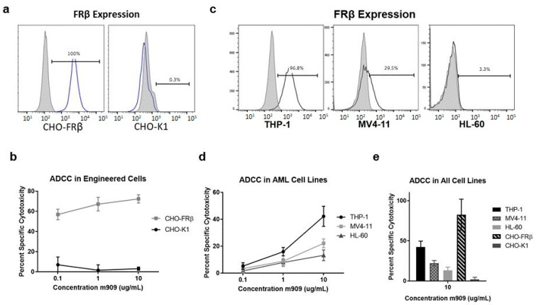
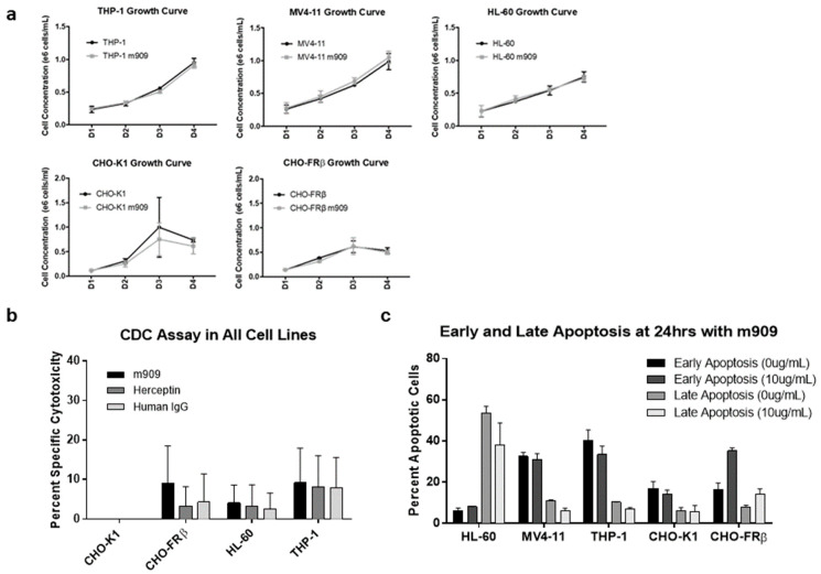
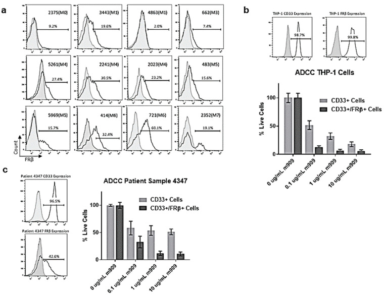
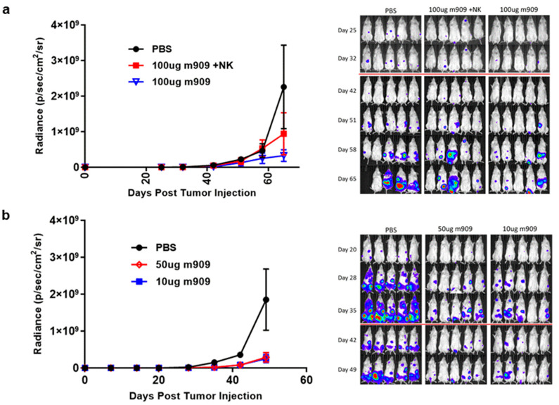
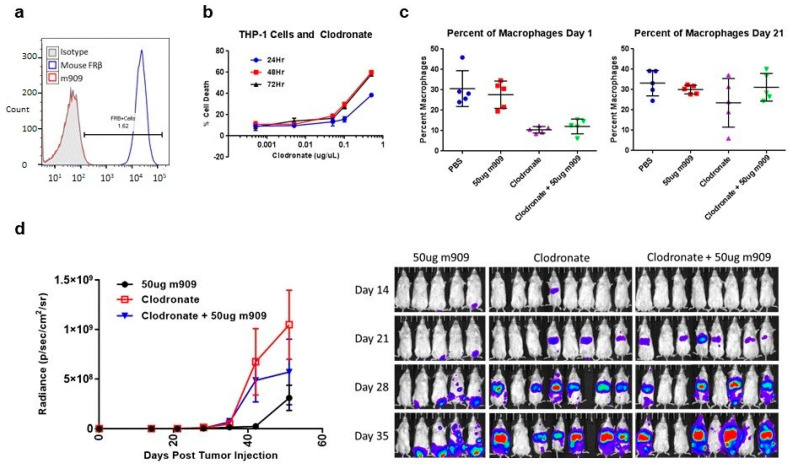
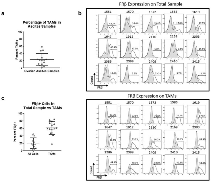
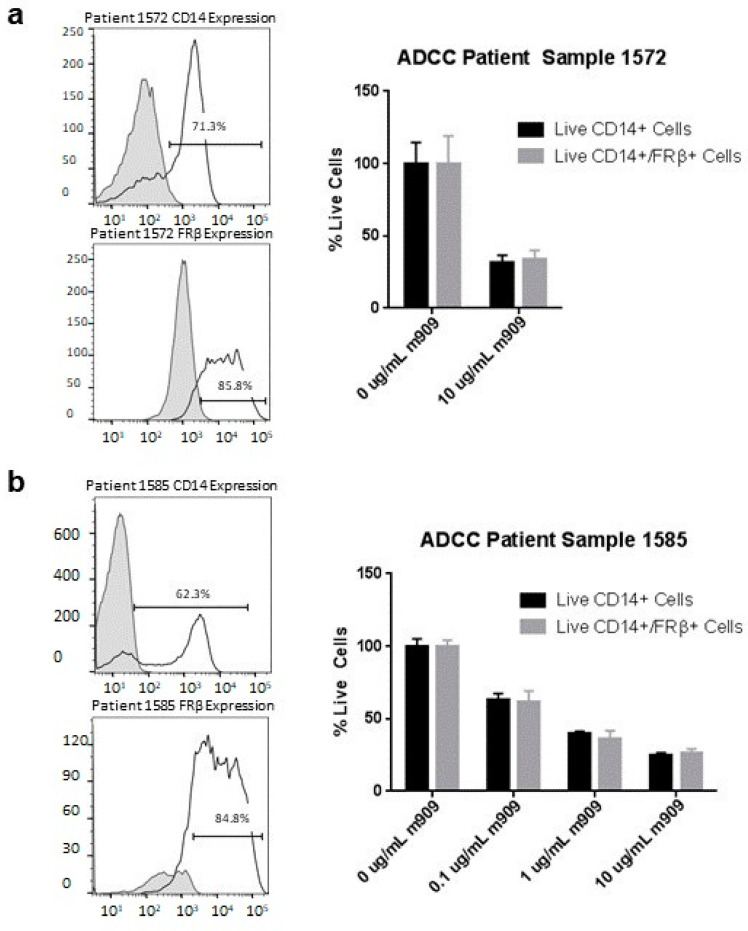
Folate receptor beta (FRβ) is a folate binding receptor expressed on myeloid lineage hematopoietic cells. FRβ is commonly expressed at high levels on malignant blasts in patients with acute myeloid leukemia (AML), as well as on M2 polarized tumor-associated macrophages (TAMs) in the tumor microenvironment of many solid tumors. Therefore, FRβ is a potential target for both direct and indirect cancer therapy. We demonstrate that FRβ is expressed in both AML cell lines and patient-derived AML samples and that a high-affinity monoclonal antibody against FRβ (m909) has the ability to cause dose- and expression-dependent ADCC against these cells in vitro. Importantly, we find that administration of m909 has a significant impact on tumor growth in a humanized mouse model of AML. Surprisingly, m909 functions in vivo with and without the infusion of human NK cells as mediators of ADCC, suggesting potential involvement of mouse macrophages as effector cells. We also found that TAMs from primary ovarian ascites samples expressed appreciable levels of FRβ and that m909 has the ability to cause ADCC in these samples. These results indicate that the targeting of FRβ using m909 has the potential to limit the outgrowth of AML in vitro and in vivo. Additionally, m909 causes cytotoxicity to TAMs in the tumor microenvironment of ovarian cancer warranting further investigation of m909 and its derivatives as therapeutic agents in patients with FRβ-expressing cancers.

Keywords: acute myeloid leukemia; folate receptor beta; ovarian cancer; tumor-associated macrophages.


Conflict of interest statement

Daniel J. Powell holds patents in the area of anti-FRβ CAR T cell therapy. No other authors have any conflicts of interest to disclose.

Figures

Figure 1
Figure 1
FRβ expression analysis and targeting using m909 antibody. (a) FRβ is expressed at 100% on the engineered cell line (CHO-FRβ). No expression is detected in the parental control (CHO-K1). (b) Antibody-dependent cytotoxicity assay (ADCC) with CHO-FRβ cells, NK cells at E:T ratio 10:1 and increasing doses of m909. Specific cytotoxicity is calculated by subtracting the percentage of cell death with no antibody. Representative results from one of two independent experiments with triplicate samples in each experiment are shown. (c) FRβ expression on immortalized AML cell lines THP-1, MV4-11, and HL-60. (d) ADCC assay at increasing doses of m909 for AML cell lines incubated with NK cells at E:T of 10:1 demonstrating a dose-dependent and expression-dependent response. Representative results from one of three independent experiments with triplicate samples per concentration and cell type are shown. (e) Specific cytotoxicity for ADCC assay for each cell line at 10 μg/mL m909 using NK cells from the same donor performed with triplicate samples. All error bars represent standard deviation.
Figure 2
Figure 2
Evaluating additional mechanisms of action for m909 antibody. (a) Cells grown in the presence or absence of 10 μg/mL of m909 over time in days. (b) Complement-dependent specific cytotoxicity for cell lines in presence of 10 μg/mL of m909, Herceptin, or human IgG1 with human serum. Samples tested in triplicate. (c) Percentage of cells with early and late markers of apoptosis after 24 h of incubation with and without 10 μg/mL m909. All error bars represent standard deviation.
Figure 3
Figure 3
Activity of m909 against primary patient AML samples. (a) FRβ expression on patient samples with various subtypes of AML. (b) Expression of CD33 and FRβ in THP-1 cells and ADCC by flow using THP-1 cells demonstrates efficacy with dose-dependent response. (c) CD33 and FRβ expression data for patient sample 4347. ADCC data for patient sample 4347 showing average from two separate experiments with triplicate samples. All graphs were normalized to wells with 0 μg/mL of m909. Error bars represent standard deviation.
Figure 4
Figure 4
Preclinical activity of m909 against THP-1 tumors in vivo. (a) Mice were injected with 2 × 10⁶ THP-1 cells expressing GFP-fLuc on day 0 and imaged weekly. They were treated with IP injection starting on day 2 every 2–3 days thereafter for 10 doses with 100 μg m909 or PBS. The NK group received activated NK cells on day 2. The average maximum radiance of each group at each measurement time point is shown in the graph. Significant differences were identified on day 65 between the treatment groups and the PBS group. (b) Mice were injected with 2 × 10⁶ THP-1 cells expressing GFP-fLuc on day 0 and received antibody via IP injection starting on day 2 and every 2–3 days thereafter at the respective doses for a total of 10 doses. Significant differences were identified between the treatment groups and the PBS group on day 49. All error bars represent standard error. Red lines on images indicate a change in exposure time.
Figure 5
Figure 5
m909 activity in vivo after macrophage depletion. (a) m909 does not cross react with mouse FRβ. (b) In vitro co-culture of THP-1 cells in the presence of liposomal clodronate at different concentrations for 24, 48, and 72 h shows increasing toxicity at higher concentrations. Error bars represent standard deviation. (c) Blood draws from five mice in each treatment group demonstrate the percentage of macrophages on day 1 and day 21. 300 μL of liposomal clodronate was given on day −1 and repeated small dose of 25μL every 5 days until the end of antibody dosing. Error bars represent standard deviation. (d) Mice injected with 2 × 10⁶ THP-1 cells on day 0 and then treated with m909 for 10 doses starting day 2 as in previous experiments as well as liposomal clodronate. m909 still has an impact on tumor growth even in the presence of liposomal clodronate. Error bars represent standard error.
Figure 6
Figure 6
Tumor associated macrophages in ovarian cancer ascites samples and expression of FRβ. (a) Percentage of TAMs making up each of the fifteen ovarian ascites samples as determined by CD11b and CD14 double-positive population with an average of 23.3%. (b) Flow plots showing the expression of FRβ in each of the 15 total ascites samples compared to expression on TAMs in the sample. (c) Comparison of the average expression of FRβ in the total sample (20.8%) versus expression on TAMs (61.6%).
Figure 7
Figure 7
Expression of FRβ and ADCC on primary ovarian ascites samples. (a) CD14 and CD14/FRβ expression data for patient sample 1572. ADCC data for patient sample 1572 showing a significant decrease in live FRβ expressing cells. (b) CD14 and CD14/FRβ expression on patient sample 1585. ADCC data for patient sample 1585 showing a dose-dependent response to m909 administration. All samples were tested in triplicate and all graphs were normalized to wells with 0 μg/mL of m909. Error bars represent standard deviation.

References

    1. Zhang H., Chen J. Current status and future directions of cancer immunotherapy. J. Cancer. 2018;9:1773–1781. doi: 10.7150/jca.24577. - DOI - PMC - PubMed
    1. Coney L.R., Tomassetti A., Carayannopoulos L., Frasca V., Kamen B.A., Colnaghi M.I., Zurawski V.R., Jr. Cloning of a tumor-associated antigen: MOv18 and MOv19 antibodies recognize a folate-binding protein. Cancer Res. 1991;51:6125–6132. - PubMed
    1. Ross J.F., Wang H., Behm F.G., Mathew P., Wu M., Booth R., Ratnam M. Folate receptor type beta is a neutrophilic lineage marker and is differentially expressed in myeloid leukemia. Cancer. 1999;85:348–357. doi: 10.1002/(SICI)1097-0142(19990115)85:2<348::AID-CNCR12>3.0.CO;2-4. - DOI - PubMed
    1. Ross J.F., Chaudhuri P.K., Ratnam M. Differential regulation of folate receptor isoforms in normal and malignant tissues in vivo and in established cell lines. Physiol. Clin. Implic. Cancer. 1994;73:2432–2443. doi: 10.1002/1097-0142(19940501)73:93.0.co;2-s. - DOI - PubMed
    1. Kandalaft L.E., Powell D.J., Coukos G., Coukos G. A phase I clinical trial of adoptive transfer of folate receptor-alpha redirected autologous T cells for recurrent ovarian cancer. J. Transl. Med. 2012;10:157. doi: 10.1186/1479-5876-10-157. - DOI - PMC - PubMed

MeSH terms