. 2021 May 25;9(6):599.
doi: 10.3390/biomedicines9060599.

Serum Amyloid A1/Toll-Like Receptor-4 Axis, an Important Link between Inflammation and Outcome of TBI Patients

Affiliations

Serum Amyloid A1/Toll-Like Receptor-4 Axis, an Important Link between Inflammation and Outcome of TBI Patients

Víctor Farré-Alins et al. Biomedicines. .

Abstract

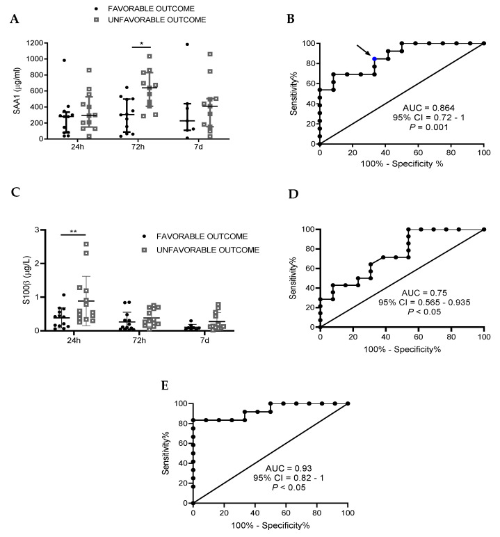
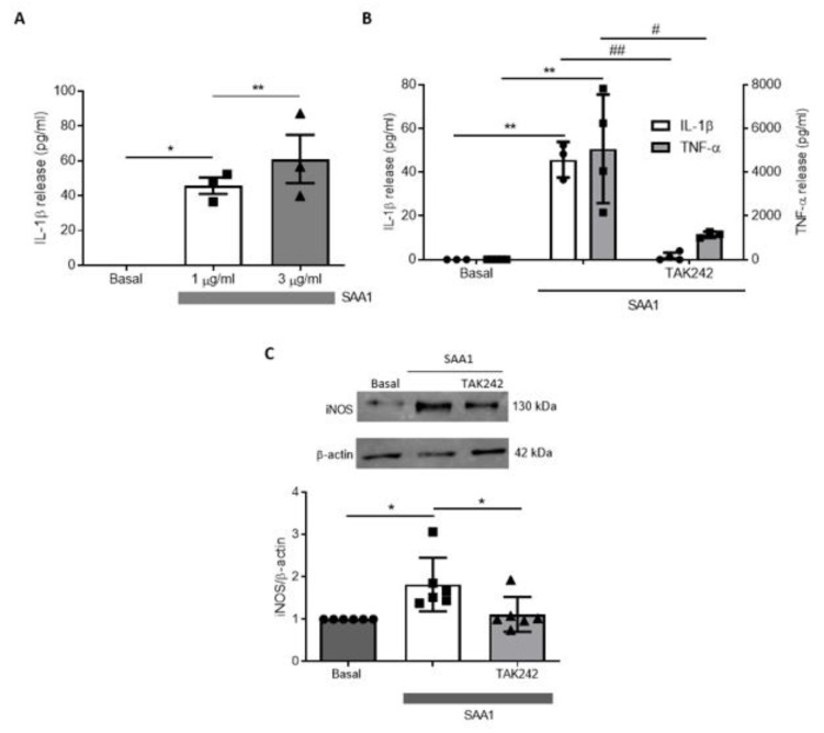
Traumatic brain injury (TBI) is one of the leading causes of mortality and disability worldwide without any validated biomarker or set of biomarkers to help the diagnosis and evaluation of the evolution/prognosis of TBI patients. To achieve this aim, a deeper knowledge of the biochemical and pathophysiological processes triggered after the trauma is essential. Here, we identified the serum amyloid A1 protein-Toll-like receptor 4 (SAA1-TLR4) axis as an important link between inflammation and the outcome of TBI patients. Using serum and mRNA from white blood cells (WBC) of TBI patients, we found a positive correlation between serum SAA1 levels and injury severity, as well as with the 6-month outcome of TBI patients. SAA1 levels also correlate with the presence of TLR4 mRNA in WBC. In vitro, we found that SAA1 contributes to inflammation via TLR4 activation that releases inflammatory cytokines, which in turn increases SAA1 levels, establishing a positive proinflammatory loop. In vivo, post-TBI treatment with the TLR4-antagonist TAK242 reduces SAA1 levels, improves neurobehavioral outcome, and prevents blood-brain barrier disruption. Our data support further evaluation of (i) post-TBI treatment in the presence of TLR4 inhibition for limiting TBI-induced damage and (ii) SAA1-TLR4 as a biomarker of injury progression in TBI patients.

Keywords: biomarkers; immunomodulation; neuroinflammation; prognosis; traumatic brain injury.


Conflict of interest statement

Some results of the study have been included in a pending patent.

Figures

Figure 1
Figure 1
SAA1 levels remains increased the first week after trauma in serum of TBI patients, while S100B just raises at 24 h. (A) SAA1 levels increased at 24 h (n = 29), 72 h (n = 28), and 7 days (n = 21) after trauma compared to healthy subjects (controls, n = 8). ** p < 0.01, *** p < 0.001, Kruskal–Wallis with Dunn’s multiple comparisons test. (B) S100B levels significantly incremented at 24 h (n = 29), but not at 72 h (n = 28) and 7 days (n = 21) after trauma. ** p < 0.01, *** p < 0.001, Kruskal–Wallis with Dunn’s multiple comparisons test. (C) Patients were classified in two groups of severity (mild and moderate/severe) according to GCS. SAA1 serum levels correlated with severity at 72 h and 1 week post-trauma. * p < 0.05, ** p < 0.01, 2-way ANOVA with Sidak’s multiple comparisons test. (D) Patients were classified in two groups of severity (mild and moderate/severe) according to GCS. S100B levels correlated with severity at 24 h. * p < 0.05, 2-way ANOVA with Sidak’s multiple comparisons test. Data of all experiments are represented as median with interquartile range.
Figure 2
Figure 2
SAA1 levels 72 h after trauma predict TBI patients’ outcome at 6 months, S100B at 24 h, and a combination of both biomarkers improves the ability of prediction. Functional outcome was assessed 6 months after trauma using the GOSE test and patients were divided in two groups: favorable and unfavorable outcome. (A) At 72 h, SAA1 levels were significantly higher in the unfavorable outcome group. * p < 0.05, 2-way ANOVA with Sidak’s multiple comparisons test. (B) Receiver operating curve (ROC) for SAA1 levels at 72 h after hospital admission to predict possible differences between “favorable” (n = 13) and “unfavorable” (n = 13) outcome. Blue dots represent the value for sensitivity and specificity shown in Table 2. (C) At 24 h, S100B in serum is increased in the unfavorable outcome group. ** p < 0.01, 2-way ANOVA with Sidak’s multiple comparisons test. (D) Receiver operating curve (ROC) for S100B levels at 24 h after hospital admission to predict possible differences between “favorable” (n = 13) and “unfavorable” (n = 13) outcome. (E) Receiver operating curve (ROC) that combines SAA1 (72 h) and S100B (24 h) values to determine the discriminative capacity of the biomarker combination in outcome 6 months after the injury (favorable, n = 13; unfavorable, n = 13). Data of 2A and 2C are represented as median with interquartile range.
Figure 3
Figure 3
TLR4 mRNA levels increase in circulating leucocytes during the first week after trauma and determination at 72 h positively correlates with SAA1 serum levels. (A) Q-PCR determined that TLR4 mRNA levels of circulating blood cells significantly increase at 24 h (n = 13) and 72 h (n = 11) in trauma patients compared to controls (n = 7). At 7 days (n = 10), there were no significant differences. * p < 0.05, 1-way ANOVA with Dunnett’s multiple comparisons test. (B) A correlation analysis (10 patients) between TLR4 mRNA of blood cells and SAA1 serum levels at 72 h was determined. The correlation coefficient (r = 0.6395) detected a positive correlation between the variables. In the correlation analysis test, p < 0.05 was established to determine a true correlation. Data of all experiments are represented as median with interquartile range.
Figure 4
Figure 4
TAK242 treatment prevented the increase of proinflammatory parameters induced by SAA1 in primary mixed glial cultures. (A) Glial cultures were treated with SAA1 (1 and 3 µg/mL) with or without TAK242 (1 µM) during 24 h. ELISA revealed a significant increase of IL-1β release to culture medium (n = 3). * p < 0.05, ** p < 0.01, 1-way ANOVA test with Tukey’s multiple comparisons test. (B) ELISA analysis revealed that TAK242 prevented the increase of IL-1β (n = 4) and TNF-α (n = 5) produced by SAA1. * p < 0.05, ** p < 0.01, # p < 0.05, ## p < 0.01,1-way ANOVA test with Tukey’s multiple comparisons test. (C) Representative immunoblot and quantification of iNOS expression in cultures treated with SAA1 with or without TAK242 (n = 6). * p < 0.05, 1-way ANOVA test with Tukey’s multiple comparisons test. Data of all experiments are represented as mean ± SD.
Figure 5
Figure 5
Glial cells synthetized and released SAA1 after inflammatory stimuli. (A) Recombinant proteins IL-1β (10 ng/mL) and TNF-α (10 ng/mL) produced the release of SAA1 (detected by ELISA) to the culture medium of mixed glial cultures (n = 6). * p < 0.05, ** p < 0.01, 2-tailed unpaired t test. (B) Primary mixed glial cultures treated with LPS 1 µg/mL stimulated SAA1 release, which was reverted by TAK242 (1 µM) (n = 5). * p < 0.05, *** p < 0.001, # p < 0.05, 1-way ANOVA test with Tukey’s multiple comparisons test. Data of all experiments are represented as mean ± SD.
Figure 6
Figure 6
TLR4 antagonism decreased serum and brain SAA1 levels 1 day after TBI. (A) TAK242 (3 mg/kg) reduced serum SAA1 protein levels. Mice were treated 1 h after CHI (sham, n = 8; vehicle, n = 10; TAK, n = 8). * p < 0.05 and *** p < 0.001, 1-way ANOVA with Tukey’s multiple comparisons test. (B) Treatment reduced cerebral accumulation of SAA1 at one day post-injury (sham, n = 4; vehicle, n = 11; TAK, n = 9). * p < 0.05, 1-way ANOVA with Tukey’s multiple comparisons test. (C) Brain lysates of mice treated with or without TAK242 were immunoblotted with SAA1 antibody and protein expression was quantified (sham, n = 4; vehicle ipsi and contra, n = 4; TAK242 ipsi and contra, n = 4). * p < 0.05, ** p < 0.01, 1-way ANOVA with Tukey’s multiple comparisons test. (D) SAA1 immunostainings and (E) quantification of SAA1 positive area of (i) and (ii) brain regions of mice subjected to TBI at the indicated time points, treated or not with TAK242 (vehicle ipsi and contra, n = 3; TAK242 ipsi and contra, n = 3). ** p < 0.01, 2-way ANOVA with Sidak’s multiple comparisons test. (F) Quantification of APP particles of (i) and (ii) regions and (G) APP immunostainings of brains of mice subjected to TBI at the indicated time points, treated or not with TAK242 (vehicle ipsi and contra, 24 h and 7 d, n = 4; TAK242 ipsi and contra, 24 h and 7 d, n = 4). *** p < 0.01, # p < 0.05, 2-way ANOVA with Sidak’s multiple comparisons test. Photos show the damaged hemisphere in an area immediately surrounding the injury site ((i) the nearest 50 µm) and in a deeper area ((ii) between 50 and 100 µm). Images are representative of each group. Scale bars: 5 µm. White arrows indicate the limit of the cortical damaged area. Data of all experiments are represented as mean ± SD. Ipsi, ipsilateral hemisphere; contra, contralateral hemisphere.
Figure 7
Figure 7
TAK242 improves NSS score and Blood–Brain Barrier (BBB) impairment produced by trauma. (A) Immediately following the 1-h NSS test, mice were treated with vehicle solution (0.9% NaCl) or TAK242 (3 mg/kg). NSS was repeated in the same mice 1 d.a.i (vehicle, n = 10; TAK, n = 8). ** p < 0.01, 2-way ANOVA with Sidak’s multiple comparisons test. (B) Mice were injected intraperitoneally with Evans Blue dye (2%) after the 1-h NSS test in vehicle and TAK242 groups and were sacrificed 24 h later. For 1 h group, it was injected immediately after TBI. For 7 d group, Evans Blue was injected the day before sacrifice. Brains were cut in 2 mm slices to analyze the stained brain surface (1 h, n = 5; vehicle, n = 7; TAK242, n = 7; 7 d, n = 6). * p < 0.05, ** p < 0.01, *** p < 0.001, 1-way ANOVA with Tukey’s multiple comparisons test. Representative photographs of the four 2-mm slices obtained in each brain are shown above the graph.

Similar articles

Cited by

References

    1. Dixon K.J. Pathophysiology of Traumatic Brain Injury. Phys. Med. Rehabil. Clin. N. Am. 2017;28:215–225. doi: 10.1016/j.pmr.2016.12.001. - DOI - PubMed
    1. Jarrahi A., Braun M., Ahluwalia M., Gupta R.V., Wilson M., Munie S., Ahluwalia P., Vender J.R., Vale F.L., Dhandapani K.M., et al. Revisiting traumatic brain injury: From molecular mechanisms to therapeutic interventions. Biomedicines. 2020;8:389. doi: 10.3390/biomedicines8100389. - DOI - PMC - PubMed
    1. Prins M., Greco T., Alexander D., Giza C.C. The pathophysiology of traumatic brain injury at a glance. DMM Dis. Model. Mech. 2013;6:1307–1315. doi: 10.1242/dmm.011585. - DOI - PMC - PubMed
    1. Capizzi A., Woo J., Verduzco-Gutierrez M. Traumatic Brain Injury: An Overview of Epidemiology, Pathophysiology, and Medical Management. Med. Clin. N. Am. 2020;104:213–238. doi: 10.1016/j.mcna.2019.11.001. - DOI - PubMed
    1. Needham E.J., Helmy A., Zanier E.R., Jones J.L., Coles A.J., Menon D.K. The immunological response to traumatic brain injury. J. Neuroimmunol. 2019;332:112–125. doi: 10.1016/j.jneuroim.2019.04.005. - DOI - PubMed

LinkOut - more resources