. 2021 May 25;10(6):1316.
doi: 10.3390/cells10061316.

The Effect of Inflammatory Priming on the Therapeutic Potential of Mesenchymal Stromal Cells for Spinal Cord Repair

Affiliations

The Effect of Inflammatory Priming on the Therapeutic Potential of Mesenchymal Stromal Cells for Spinal Cord Repair

Inés Maldonado-Lasunción et al. Cells. .

Abstract

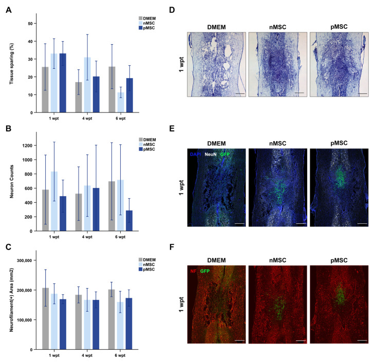
Mesenchymal stromal cells (MSC) are used for cell therapy for spinal cord injury (SCI) because of their ability to support tissue repair by paracrine signaling. Preclinical and clinical research testing MSC transplants for SCI have revealed limited success, which warrants the exploration of strategies to improve their therapeutic efficacy. MSC are sensitive to the microenvironment and their secretome can be altered in vitro by exposure to different culture media. Priming MSC with inflammatory stimuli increases the expression and secretion of reparative molecules. We studied the effect of macrophage-derived inflammation priming on MSC transplants and of primed MSC (pMSC) acute transplants (3 days) on spinal cord repair using an adult rat model of moderate-severe contusive SCI. We found a decrease in long-term survival of pMSC transplants compared with unprimed MSC transplants. With a pMSC transplant, we found significantly more anti-inflammatory macrophages in the contusion at 4 weeks post transplantation (wpt). Blood vessel presence and maturation in the contusion at 1 wpt was similar in rats that received pMSC or untreated MSC. Nervous tissue sparing and functional recovery were similar across groups. Our results indicate that macrophage-derived inflammation priming does not increase the overall therapeutic potential of an MSC transplant in the adult rat contused spinal cord.

Keywords: MSC; SCI; angiogenesis; cell therapy; contusion; immunomodulation; macrophages; neuroprotection; repair.


Conflict of interest statement

The authors declare no conflict of interest.

Figures

Figure 1
Figure 1
Experimental design. (A) The bone marrow was extracted from rats’ femurs and tibias and cultured on plastic dishes for 24 h. Then, monocytes were sorted using FACS and cultured for 7 days until differentiated to bone-marrow-derived macrophages. Mature macrophages were polarized to pro-inflammatory cells by culture in medium with LPS and IFNγ for 24 h. The conditioned medium (CM) from pro-inflammatory macrophages was collected and, later, used to prime MSC. In parallel, bone-marrow-derived MSC, adhered to the dish during the 24 h of bone marrow culture, were maintained in culture until passage 2, then FACS-sorted for purification. Sorted MSC were transduced with GFP-lentivirus to turn them green and trackable. P4 GFP-positive MSC were divided into two batches; one was cultured for 24 h in pro-inflammatory macrophage CM (primed MSC, pMSC) and the other one was cultured for 24 h in fresh D10 (naïve MSC, nMSC). pMSC and nMSC were trypsinized and resuspended in DMEM:F12 for transplantation. (B) Rats were acclimated to the behavioral tests before surgery, and the baseline scores for each test were obtained. Surgery for contusive SCI was performed. and the next day, BBB scores of the injured rats were collected. Three days after injury, surgery for direct injection of pMSC, nMSC, or DMEM:F12 was performed. At 1 and 4 wpt, six rats per group and time point were fixed and their spinal cords dissected for histological analysis. Ten additional rats per group were kept alive for 6 wpt, and their sensorimotor performance was measured using BBB scores weekly after transplant, the horizontal ladder test at 3 and 6 wpt, and the Von Frey test at 4 and 6 wpt. At 6 wpt, those rats were fixed and their spinal cords dissected for histological analysis. Abbreviations: MSC: mesenchymal stromal cell; P2: passage 2; LPS: lipopolysaccharide; IFNγ: interferon-gamma; GFP: green fluorescent protein; LV: lentivirus; D10: DMEM medium with 10% fetal bovine serum and 0.1% gentamycin; SCI: spinal cord injury; BBB: Basso, Beattie, Bresnahan locomotor scale; wpt: weeks post-transplant.
Figure 2
Figure 2
Inflammatory priming decreases long-term survival of MSC in the contused spinal cord. (A) Photomicrographs representing examples of injured spinal cords at 1 and 6 wpt for the vehicle injected animals (DMEM), the nMSC transplanted animals, and the pMSC transplanted animals. The immunostaining marks astrocytes in red (GFAP), transplants in green (GFP), and cell nuclei in blue (DAPI). The dashed lines represent the manually selected regions of interest (ROIs) to designate the injury cavity. Any green fluorescence detected on the DMEM images is a product of auto fluorescence, since those animals did not receive LV-GFP transduced cells. Scale bars represent 500 µm (Overview: 4×, image scan: 20×). The spinal cords’ orientation is top–rostral and bottom–caudal. Intensity has been equally modified on all images for illustrative purposes only but not for quantitative analysis. (B) Bar graph representing the percentage of the injury cavity occupied by GFP-positive area over time from those animals that received a cell transplant. pMSC occupy eight-fold less area than nMSC at 6 wpt. The DMEM group was not included in the graph because the values are negligible. The bars represent mean and SEM. n = 6 per group and time point. Statistical significance (noted on the graph) was accepted at p < 0.05. Abbreviations: DMEM: Dubbelco’s Modified Eagle’s Medium; nMSC: naïve MSC; pMSC: primed MSC; wpt: weeks post-transplant; GFP: green fluorescent protein; MSC: mesenchymal stromal cell; GFAP: glial fibrillary acidic protein.
Figure 3
Figure 3
All experimental groups have similar effects on nervous tissue sparing. (A) Bar graph representing the percentage of healthy tissue spared, relative to an equally sized healthy spinal cord segment over time. No significant differences were found between groups. (B) Bar graph representing the number of neurons present just rostral and caudal to the injury over time. No significant differences were found between groups. (C) Bar graph representing axonal presence in the injury over time. No significant differences were found between groups. On (AC), bars represent the mean and SEM. n = 6 per group and time point. (DF) Example photomicrographs of injured spinal cords at 1 wpt are shown for visualization purposes. The spinal cords’ orientation is top–rostral and bottom–caudal. (D) Tissue sparing was quantified using cresyl violet staining imaged under a bright field microscope (2.5× magnification). (E) Neuronal bodies were quantified using NeuN (white), GFP (green), and DAPI (blue) staining. (F) Axonal presence was quantified using NF (red) and GFP (green) staining. For (E,F), intensity was equally modified on all images for illustrative purposes only but not for quantitative analysis (Overview: 4×, image scan: 20×). Any green fluorescence detected on the DMEM images on E and F is product of auto fluorescence since those animals did not receive LV-GFP transduced cells. Scale bars on all images represent 500 µm. Abbreviations: DMEM: Dubbelco’s Modified Eagle’s Medium; nMSC: naïve MSC; pMSC: primed MSC; wpt: weeks post-transplant; GFP: green fluorescent protein; MSC: mesenchymal stromal cell; NeuN: neuronal nuclei; NF: neurofilament.
Figure 4
Figure 4
pMSC transplants induce transient immunomodulation at 4 wpt. (AC) Example photomicrographs of injured spinal cords at 4 wpt. Inlets represent a close-up detail of the staining; a square on the overview designates the source for the inlet image. The spinal cords’ orientation is top–rostral and bottom–caudal. (A) Pro-inflammatory macrophages were quantified using CD86 (red), ED1 (white), and DAPI (blue) staining. (B) Early-acting pro-inflammatory macrophages were quantified using CD206 (red), ED1 (white), and DAPI (blue) staining. (C) Late-acting pro-inflammatory macrophages were quantified using CD163 (red), ED1 (white), and DAPI (blue) staining. For (AC), intensity was equally modified on all images for illustrative purposes only but not for quantitative analysis. Scale bars on all images represent 500 µm for the overviews and 25 µm for the inlets (Overviews: 4×, image scan: 20×, Inlets: 20×). (D) Bar graph representing the percentage of pro-inflammatory macrophages, relative to the total number of macrophages in the injury at 4 wpt. No significant differences were found between groups. (E) Bar graph representing the percentage of early anti-inflammatory macrophages, relative to the total number of macrophages in the injury at 4 wpt. pMSC transplants result in 20% more CD206-positive macrophages compared to controls. (F) Bar graph representing the percentage of late anti-inflammatory macrophages, relative to the total number of macrophages in the injury at 4 wpt. No significant differences were found between groups. On (DF), bars represent the mean and SEM, and significance was accepted at p < 0.05. n = 6 per group and time point. Abbreviations: DMEM: Dubbelco’s Modified Eagle’s Medium; nMSC: naïve MSC; pMSC: primed MSC; wpt: weeks post-transplant; MSC: mesenchymal stromal cell.
Figure 5
Figure 5
pMSC and nMSC transplants promote angiogenesis at 1 wpt. (AC) Example photomicrographs of injured spinal cords at 1 wpt. The spinal cords’ orientation is top–rostral and bottom–caudal. (A) Blood vessel presence in the injury was measured using RECA-1 (red) staining. (B) Blood vessel maturation in the injury was measured using occludin (red) staining, and (C). ZO-1 (white) staining. For (AC), intensity was equally modified on all images for illustrative purposes only but not for quantitative analysis. Scale bars on all images represent 500 µm (Overviews: 4×, image scan: 20×). (D) Bar graph representing the area of RECA-1 in the epicenter of the injury over time. nMSC transplants result in almost three-fold more blood vessel presence compared to controls at 1 wpt. (E) Bar graph representing the area of occludin in the injury epicenter over time. nMSC and pMSC transplants result in two-fold more occludin presence compared to controls at 1 wpt. (F) Bar graph representing the area of ZO-1 in the injury epicenter at 1 wpt. No significant differences were found between groups. In (DF), bars represent the mean and SEM, and significance was accepted at p < 0.05. n = 6 per group and time point. Abbreviations: DMEM: Dubbelco’s Modified Eagle’s Medium; nMSC: naïve MSC; pMSC: primed MSC; wpt: weeks post-transplant; MSC: mesenchymal stromal cell; RECA-1: rat endothelial cell antigen 1; ZO-1: zonula occludens 1.

Similar articles

Cited by

References

    1. Drago D., Cossetti C., Iraci N., Gaude E., Musco G., Bachi A., Pluchino S. The stem cell secretome and its role in brain repair. Biochimie. 2013;95:2271–2285. doi: 10.1016/j.biochi.2013.06.020. - DOI - PMC - PubMed
    1. Johnson T.V., DeKorver N.W., Levasseur V.A., Osborne A., Tassoni A., Lorber B., Heller J.P., Villasmil R., Bull N.D., Martin K.R., et al. Identification of retinal ganglion cell neuroprotection conferred by platelet-derived growth factor through analysis of the mesenchymal stem cell secretome. Brain. 2014;137:503–519. doi: 10.1093/brain/awt292. - DOI - PMC - PubMed
    1. Asami T., Ishii M., Fujii H., Namkoong H., Tasaka S., Matsushita K., Ishii K., Yagi K., Fujiwara H., Funatsu Y., et al. Modulation of murine macrophage TLR7/8-mediated cytokine expression by mesenchymal stem cell-conditioned medium. Mediat. Inflamm. 2013;2013:264260. doi: 10.1155/2013/264260. - DOI - PMC - PubMed
    1. Sun G., Li G., Li D., Huang W., Zhang R., Zhang H., Duan Y., Wang B. hucMSC derived exosomes promote functional recovery in spinal cord injury mice via attenuating inflammation. Mater. Sci. Eng. C Mater. Biol. Appl. 2018;89:194–204. doi: 10.1016/j.msec.2018.04.006. - DOI - PubMed
    1. Liu Y., Dulchavsky D.S., Gao X., Kwon D., Chopp M., Dulchavsky S., Gautam S.C. Wound repair by bone marrow stromal cells through growth factor production. J. Surg. Res. 2006;136:336–341. doi: 10.1016/j.jss.2006.07.037. - DOI - PubMed

Publication types