Review
. 2021 May 25;10(6):1317.
doi: 10.3390/cells10061317.

Mitochondrial Ca2+ Signaling in Health, Disease and Therapy

Affiliations
Review

Mitochondrial Ca2+ Signaling in Health, Disease and Therapy

Lorenzo Modesti et al. Cells. .

Abstract

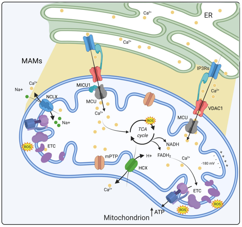
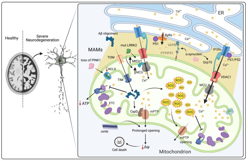
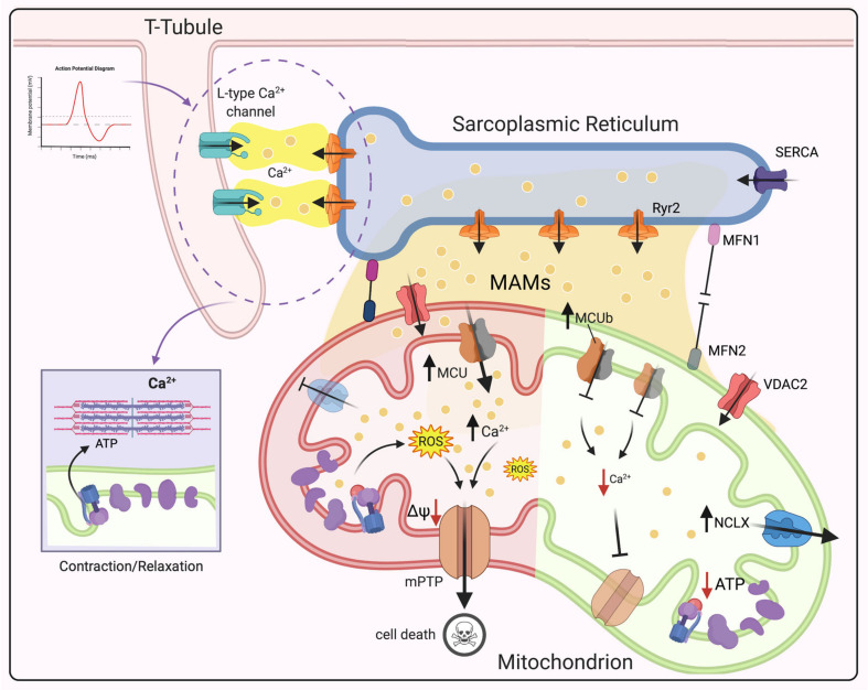
The divalent cation calcium (Ca2+) is considered one of the main second messengers inside cells and acts as the most prominent signal in a plethora of biological processes. Its homeostasis is guaranteed by an intricate and complex system of channels, pumps, and exchangers. In this context, by regulating cellular Ca2+ levels, mitochondria control both the uptake and release of Ca2+. Therefore, at the mitochondrial level, Ca2+ plays a dual role, participating in both vital physiological processes (ATP production and regulation of mitochondrial metabolism) and pathophysiological processes (cell death, cancer progression and metastasis). Hence, it is not surprising that alterations in mitochondrial Ca2+ (mCa2+) pathways or mutations in Ca2+ transporters affect the activities and functions of the entire cell. Indeed, it is widely recognized that dysregulation of mCa2+ signaling leads to various pathological scenarios, including cancer, neurological defects and cardiovascular diseases (CVDs). This review summarizes the current knowledge on the regulation of mCa2+ homeostasis, the related mechanisms and the significance of this regulation in physiology and human diseases. We also highlight strategies aimed at remedying mCa2+ dysregulation as promising therapeutical approaches.

Keywords: Ca2+; cancer; cardiovascular diseases; mPTP; mitochondria; neurodegenerative diseases; therapy.


Conflict of interest statement

The authors declare no conflict of interests.

Figures

Figure 1
Figure 1
Mitochondrial calcium homeostasis. mCa2+ homeostasis is tightly regulated by influx and efflux mechanisms. Ca2+ enters into the mitochondrial matrix via MCU and through a high electronegative potential (−180 mV) while its extrusion depends on NCLX and HCX exchangers. Within the matrix, Ca2+ stimulates the activity of three dehydrogenases of the Krebs cycle and ATP production. Ca2+ ions are depicted as yellow dots. Abbreviations: ER, endoplasmic reticulum; MAMs, mitochondria associated membranes; ETC, electron transport chain; MCU, mitochondrial calcium uniporter; VDAC1, voltage-dependent anion channel 1; ATP, adenosine triphosphate; MICU1, mitochondrial calcium uptake 1; IP3Rs, inositol-1,4,5-trisphosphate receptors; ROS, reactive oxygen species; mPTP, mitochondrial permeability transition pore; NCLX, Na+/Ca2+ exchanger; HCX, H+/Ca2+ exchanger (Created with Biorender.com).
Figure 2
Figure 2
Schematic view of the role of Ca2+ dysregulation in cancer and the cell cycle with a particular focus on alterations in mCa2+ levels. Ca2+-regulating proteins and channels are involved in cell cycle progression, and their dysregulation leads to alterations in the cell cycle. STIM1/ORAI1-mediated augmentation of SOCE promotes tumor growth and metastasis; VGCCs are involved in cell proliferation regulation, and T-type channel inhibition provokes cell cycle arrest followed by a significant increase in the number of cells in G1 phase and a decrease in the number of cells in S phase; TRPC1 inhibition reduces the adhesion, invasion and proliferation of different cancer cell lines and G(0)/G(1) cell cycle arrest of glioma and lung carcinoma cell lines. MCU silencing inhibits cell migration and invasion without affecting proliferation rates or apoptosis levels. MCUb silencing limits proliferation, migration, and invasion, as well as glioma progression in vivo. Loss of VDAC1 leads to ATP depletion, which results in decreased cell growth and migration in several cancer cell lines both in vitro and in vivo. The interaction between VDAC and Bcl-XL is responsible for increased ATP production in breast cancer cells, which leads to an increased migration rate. AKT-mediated phosphorylation of MICU1 causes an increase in mCa2+ content under basal conditions and ROS production, which promotes AKT-mediated tumor growth. At MAMs, IP3R3 FBXL2-dependent degradation is enhanced in cancer cells upon loss of PTEN, resulting in apoptosis resistance. The tumor suppressor BAP1 is capable of binding, deubiquitylating and stabilizing IP3R3 channels, modulating Ca2+ release into the cytosol and then into mitochondria and thus promoting cell death. Abbreviations: SOCE, store-operated calcium entry; VGCCs, voltage-gated calcium channels; TRPC1, transient receptor potential channel 1; MCU, mitochondrial calcium uniporter, MCUb, mitochondrial calcium uniporter b subunit; VDAC1, voltage-dependent anion channel 1; ATP, adenosine triphosphate; Bcl-XL, B-cell lymphoma XL; AKT, protein kinase B; MICU1, mitochondrial calcium uptake 1; IP3R3, type 3 inositol-1,4,5-trisphosphate receptor; FBXL2, F-box and leucine-rich repeat protein 2; BAP1, BRCA1-associated protein 1; PTEN, phosphatase and tensin homolog; ROS, Reactive oxygen species (Created with Biorender.com).
Figure 3
Figure 3
Schematic view of the role of mCa2+ dysregulation in neurodegenerative diseases. In AD, Aβ oligomers enter mitochondria via the translocases TOM and TIM. Once inside the matrix, they interact with specific intramitochondrial targets: (i) respiratory chain complexes III and IV, leading to ATP synthesis reduction, and (ii) CypD, leading to mPTP opening, ΔΨm collapse and activation of cell death. Excessive levels of mCa2+ can enhance ROS production, reduce the ΔΨm, and stimulate mPTP opening, thus leading to the release of proapoptotic factors. In HD, mHtt decreases the Ca2+ threshold necessary to trigger mPTP opening, preventing the binding of mPTP inhibitors and consequently augmenting its activation by increasing the binding affinity of CypD and Ca2+. In PD, α-synuclein interacts with the chaperone Grp75, thus contributing to enhancement of ER–mitochondria communication. Overexpression of wt and mutant α-synuclein leads to the destruction of VAPB-PTPIP51 tethers through its bond with VAPB, causing a decrease in the ER–mitochondria association. PINK1 deficiency results in mitochondrial calcium overload and subsequent ROS production due to negative regulation of NCLX. Mutant LRRK2 induces transcriptional upregulation of MCU and MICU1, thus leading to mCa2+ accumulation. Mutant PS1 and PS2 interact and modulate the IP3R Ca2+ release channel causing a strong stimulatory effect on its gating activity. PS1 N-terminal region can interact with ryanodine receptor (RyR) and enhance its activity. Abbreviations: ER, endoplasmic reticulum; MAMs, mitochondria-associated membranes; mPTP, mitochondrial permeability transition pore; CypD, cyclophilin D; ROS, reactive oxygen species; TIM, translocase of the inner membrane; TOM, translocase of the outer membrane; MICU1, mitochondrial calcium uptake 1; MCU, mitochondrial calcium uptake; NCLX, Na+/Ca2+ exchanger; Grp75, glucose-regulated protein 75; IP3R3, inositol-1,4,5-trisphosphate receptor type 3; ETC, electron transport chain; PINK1, PTEN-induced kinase 1; LRRK2, leucine-rich repeat kinase 2; mHtt, mutant Htt; ΔΨm, mitochondrial membrane potential; PS1, presenilin 1; PS2, presenilin 2 (Created with Biorender.com).
Figure 4
Figure 4
Schematic of the role of mCa2+ dysregulation in CVDs. Under physiological conditions, after sarcolemma depolarization, the opening of L-type Ca2+ channels allow Ca2+ to enter the cell, which stimulates Ca2+ release from the SR via RyR2. Then, Ca2+ binds to troponin C, leading to cardiomyocyte contraction. Mitochondria are juxtaposed to the SR, and high-Ca2+ microdomains form at this interface. Ca2+-mediated crosstalk between the SR and mitochondria is mediated by RyR2 and VDAC2. Reduced mCa2+ uptake by MCU deletion leads to inhibition of mPTP opening. MCUb overexpression causes a reduction in mCa2+ uptake and consequent inhibition of mPTP opening after reperfusion. Increased NCLX activity leads to Krebs cycle impairment, bioenergetic dysfunction, a decrease in NADPH levels and ROS hyperproduction, while NCLX knockout triggers mCa2+ overload and consequent mPTP opening. Ca2+ leakage through RyR2 on the SR causes mCa2+ accumulation and ROS production, leading to chronic HF after mPTP opening. MFN2 ablation may reduce mPTP opening by decreasing mCa2+ uptake, thus protecting the heart from IRI. Deletion of both MFN1 and MFN2 in adult hearts induces lethal dilated cardiomyopathy. Abbreviations: MAMs, Mitochondria-associated membranes; SR, sarcoplasmic reticulum; RyR2, ryanodine receptor type 2; SERCA, Sarco-Endoplasmic Reticulum Calcium ATPase; MCU, mitochondrial calcium uniporter; VDAC2, voltage-dependent anion channel 2; mPTP, mitochondrial permeability transition pore; NCLX, Na+/Ca2+ exchanger; ROS, reactive oxygen species; ΔΨm, mitochondrial membrane potential; MFN1, mitofusin 1; MFN2, mitofusin 2; (Created with Biorender.com).

References

    1. Giorgi C., Danese A., Missiroli S., Patergnani S., Pinton P. Calcium Dynamics as a Machine for Decoding Signals. Trends Cell Biol. 2018;28:258–273. doi: 10.1016/j.tcb.2018.01.002. - DOI - PubMed
    1. Vakifahmetoglu-Norberg H., Ouchida A.T., Norberg E. The role of mitochondria in metabolism and cell death. Biochem. Biophys. Res. Commun. 2017;482:426–431. doi: 10.1016/j.bbrc.2016.11.088. - DOI - PubMed
    1. Ma K., Chen G., Li W., Kepp O., Zhu Y., Chen Q. Mitophagy, Mitochondrial Homeostasis, and Cell Fate. Front. Cell Dev. Biol. 2020;8:467. doi: 10.3389/fcell.2020.00467. - DOI - PMC - PubMed
    1. Yapa N.M.B., Lisnyak V., Reljic B., Ryan M.T. Mitochondrial dynamics in health and disease. FEBS Lett. 2021;595:1184–1204. doi: 10.1002/1873-3468.14077. - DOI - PubMed
    1. Clapham D.E. Calcium signaling. Cell. 2007;131:1047–1058. doi: 10.1016/j.cell.2007.11.028. - DOI - PubMed

Publication types

LinkOut - more resources