. 2021 May 30;22(11):5859.
doi: 10.3390/ijms22115859.

Regulation of ClC-2 Chloride Channel Proteostasis by Molecular Chaperones: Correction of Leukodystrophy-Associated Defect

Affiliations

Regulation of ClC-2 Chloride Channel Proteostasis by Molecular Chaperones: Correction of Leukodystrophy-Associated Defect

Ssu-Ju Fu et al. Int J Mol Sci. .

Abstract

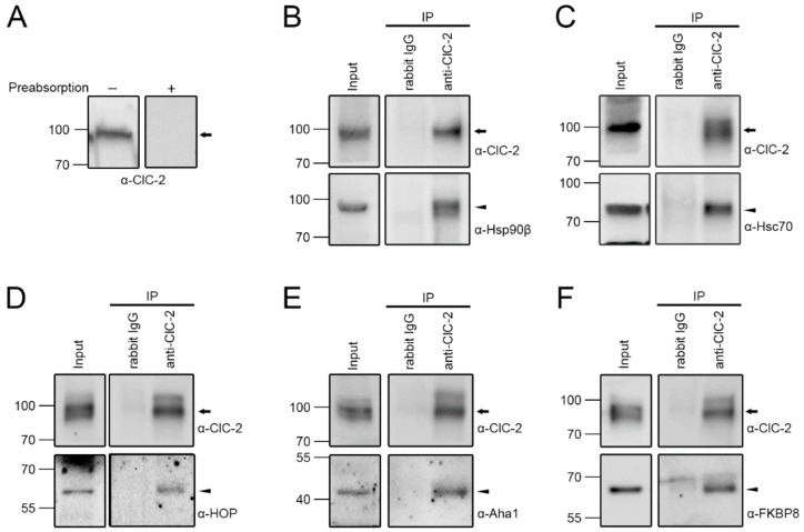
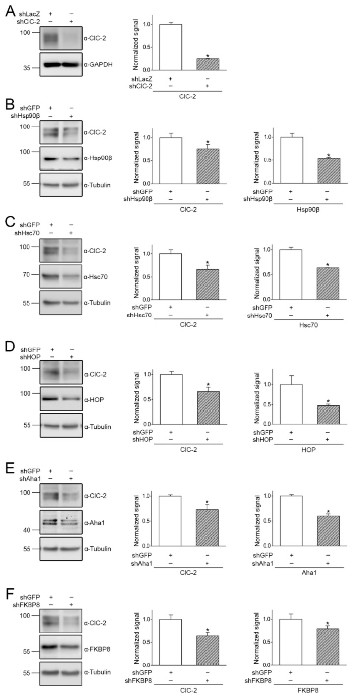
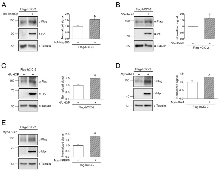
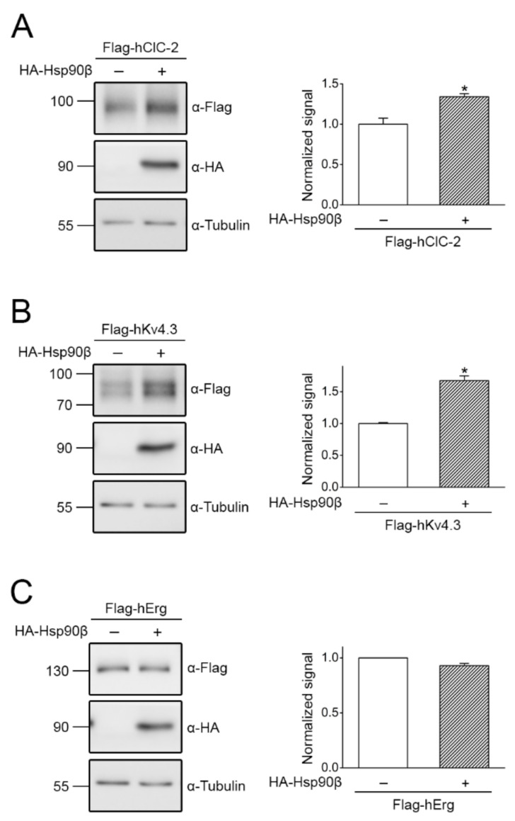
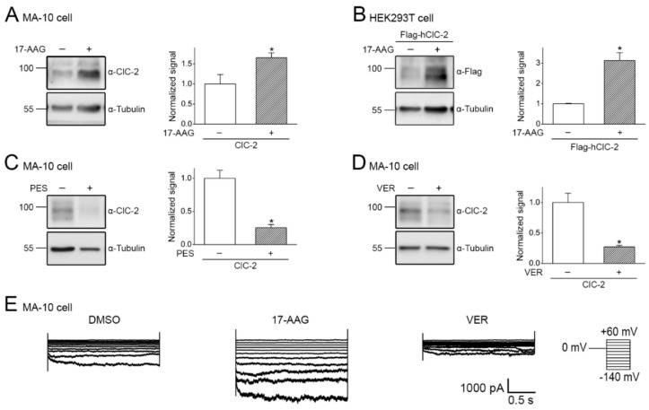
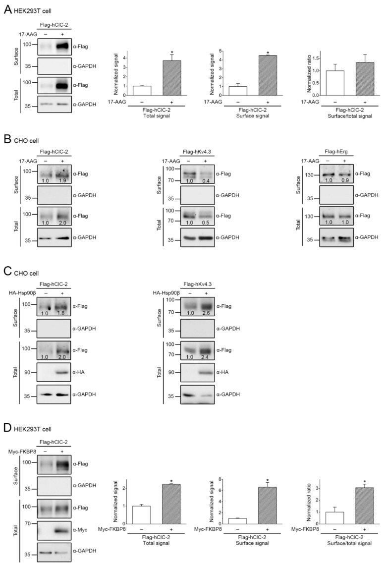
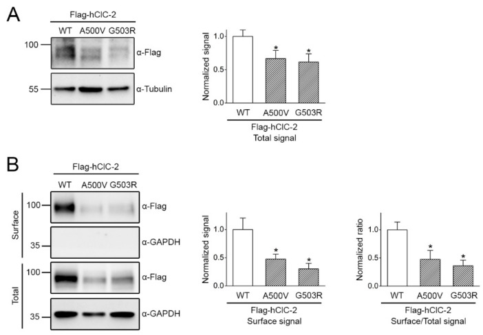
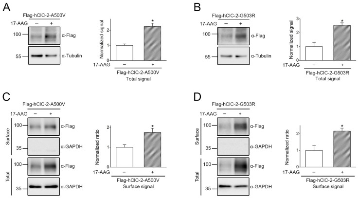
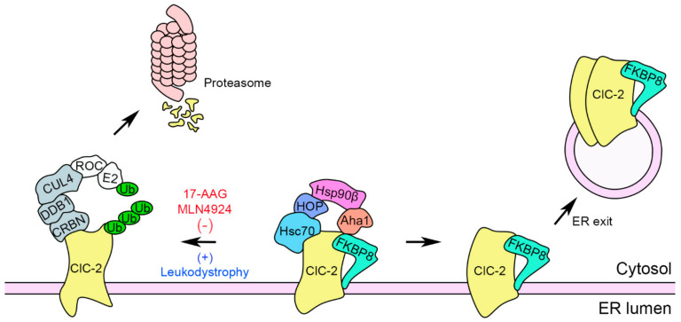
The ClC-2 channel plays a critical role in maintaining ion homeostasis in the brain and the testis. Loss-of-function mutations in the ClC-2-encoding human CLCN2 gene are linked to the white matter disease leukodystrophy. Clcn2-deficient mice display neuronal myelin vacuolation and testicular degeneration. Leukodystrophy-causing ClC-2 mutant channels are associated with anomalous proteostasis manifesting enhanced endoplasmic reticulum (ER)-associated degradation. The molecular nature of the ER quality control system for ClC-2 protein remains elusive. In mouse testicular tissues and Leydig cells, we demonstrated that endogenous ClC-2 co-existed in the same protein complex with the molecular chaperones heat shock protein 90β (Hsp90β) and heat shock cognate protein (Hsc70), as well as the associated co-chaperones Hsp70/Hsp90 organizing protein (HOP), activator of Hsp90 ATPase homolog 1 (Aha1), and FK506-binding protein 8 (FKBP8). Further biochemical analyses revealed that the Hsp90β-Hsc70 chaperone/co-chaperone system promoted mouse and human ClC-2 protein biogenesis. FKBP8 additionally facilitated membrane trafficking of ClC-2 channels. Interestingly, treatment with the Hsp90-targeting small molecule 17-allylamino-17-demethoxygeldanamycin (17-AAG) substantially boosted ClC-2 protein expression. Also, 17-AAG effectively increased both total and cell surface protein levels of leukodystrophy-causing loss-of-function ClC-2 mutant channels. Our findings highlight the therapeutic potential of 17-AAG in correcting anomalous ClC-2 proteostasis associated with leukodystrophy.

Keywords: 17-AAG; channelopathy; chaperone; co-chaperone; protein quality control; proteostasis.


Conflict of interest statement

The authors declare no competing financial interests.

Figures

Figure 1
Figure 1
Interaction of molecular chaperones and co-chaperones with endogenous ClC-2 in mouse testes. (A) Endogenous ClC-2 protein signal in mouse testes. The specificity of the rabbit-derived anti-ClC-2 antibody (α-ClC-2) was verified by preabsorption with a control antigen peptide. (BF) Co-immunoprecipitation of endogenous Hsp90β (B), Hsc70 (C), HOP (D), Aha1 (E), or FKBP8 (F) with ClC-2. Mouse testis lysates were immunoprecipitated (IP) with the rabbit IgG or α-ClC-2. The molecular weight markers (in kilodaltons) and immunoblotting antibodies (α-Hsp90β, α-Hsc70, α-HOP, α-Aha1, and α-FKBP8) are labeled to the left and right, respectively. The protein bands corresponding to endogenous ClC-2 and chaperones/co-chaperones are highlighted with the black arrow and the black arrowhead, respectively. Corresponding expression levels of ClC-2 and chaperones/co-chaperones in the lysates are shown in the Input lane. In all cases hereafter, input represents about 10% of the total protein used for immunoprecipitation.
Figure 2
Figure 2
Co-localization of molecular chaperones and co-chaperones with endogenous ClC-2 in mouse MA-10 cells. (A) (Left) Endogenous ClC-2 protein signal in mouse MA-10 Leydig cells. The specificity of the mouse-derived anti-ClC-2 antibody was verified by shRNA knock-down of mouse ClC-2 (shClC-2). shRNA knock-down of LacZ (shLacZ) was employed as the control experiment. GAPDH was used as the loading control. (Right) Quantification of relative ClC-2 protein levels. Protein density was standardized as the ratio of the ClC-2 signal to the cognate GAPDH signal. Values from the shClC-2 group (hatched bar) was then normalized to those for the corresponding shLacZ control (clear bar). Asterisk denotes significant difference from the shLacZ control (*, t test: p < 0.05; n = 4). (BF) Representative confocal micrographs showing the immunofluorescence staining patterns of endogenous ClC-2 (green) and chaperones/co-chaperones (red). Fixed MA-10 cells were stained with the mouse-derived anti-ClC-2 antibody, as well as rabbit-derived antibodies against the indicated molecular chaperones/co-chaperones, under the permeabilized configuration. Merged images of ClC-2 and chaperone/co-chaperone signals are shown in the rightmost panels, where DAPI (blue) was employed as a nuclear counterstain. Arrowheads denote plasma membrane-localization of ClC-2. Scale bar = 15 μm. Data are representative of at least three independent experiments.
Figure 3
Figure 3
shRNA knock-down of endogenous chaperones or co-chaperones in mouse MA-10 cells. (A) Verification of the specificity of the rabbit-derived anti-ClC-2 antibody in mouse MA-10 Leydig cells. (Left) Representative immunoblot showing the effect of shLacZ and shClC-2 knock-down on endogenous ClC-2 protein signal. (Right) Quantification of relative ClC-2 protein levels. Protein density was standardized as the ratio of the ClC-2 signal to the cognate GAPDH signal. Values from the shClC-2 group (hatched bar) was then normalized to those for the corresponding shLacZ control (clear bar). Asterisk denotes significant difference from the shLacZ control (*, t test: p < 0.05; n = 3). (BF) (Left panels) Representative immunoblots showing the effect of shRNA knock-down of chaperones/co-chaperones (shHsp90β, shHsc70, shHOP, shAha1, and shFKBP8) on endogenous ClC-2 expression. Infection with shGFP was used as the control experiment. Tubulin was used as the loading control. (Right panels) Quantification of relative ClC-2 and chaperone/co-chaperone protein levels. Protein density was standardized as the ratio of the ClC-2 signal to the cognate tubulin signal. Values from the shRNA knock-down groups (hatched bars) were then normalized to those for the corresponding shGFP control (clear bars). Asterisks denote significant difference from the shGFP control (*, t test: p < 0.05; n = 4–10).
Figure 4
Figure 4
Co-expression with chaperone/co-chaperone increases human ClC-2 protein level in HEK293T cells. Heterologous expression of Flag-tagged human ClC-2 channels (Flag-hClC-2) in HEK293T cells. (Left panels) Representative immunoblots showing the effect of co-expressing HA-tagged Hsp90β (HA-Hsp90β) (A), V5-tagged Hsc70 (V5-Hsc70) (B), HA-tagged HOP (HA-HOP) (C), Myc-tagged Aha1 (Myc-Aha1) (D), or Myc-tagged FKBP8 (Myc-FKBP8) (E) on Flag-hClC-2 protein expression. cDNA for Flag-hClC-2 was co-transfected with that for the indicated chaperone/co-chaperone in the molar ratio 1:3. Co-expression with the empty vector was employed as the control experiment. Tubulin was used as the loading control. (Right panels) Quantification of relative ClC-2 protein levels in the presence of various chaperones/co-chaperones. Protein density was standardized as the ratio of the ClC-2 signal to the cognate tubulin signal. Values from the chaperone/co-chaperone co-expression groups (hatched bars) were then normalized to those for the corresponding vector control (clear bars). Asterisks denote significant difference from the vector control (*, t test: p < 0.05; n = 5–11).
Figure 5
Figure 5
The effect of Hsp90β co-expression on human ClC-2, KV4.3, and Erg channel protein levels in CHO cells. Heterologous expression of Flag-hClC-2 (A), Flag-tagged human KV4.3 (Flag-hKV4.3) (B), or Flag-tagged human Erg (Flag-hErg) (C) channels in CHO cells. (Left panels) Representative immunoblots showing the effect of HA-Hsp90β co-expression on total protein levels. cDNA for Flag-hClC-2/hKV4.3/hErg was co-transfected with that for HA-Hsp90β in the molar ratio 1:3. Co-expression with the empty vector was employed as the control experiment. Tubulin was used as the loading control. (Right panels) Quantification of relative total protein levels in response to HA-Hsp90β co-expression. Protein density was standardized as the ratio of the ClC-2/KV4.3/Erg signal to the cognate tubulin signal. Values from the HA-Hsp90β co-expression groups (hatched bars) were then normalized to those for the corresponding vector control (clear bars). Asterisks denote significant difference from the vector control (*, t test: p < 0.05; n = 3).
Figure 6
Figure 6
17-AAG enhances total ClC-2 protein expression. (A,B) The effect of 24-h treatment of 1 μM 17-AAG (in 0.1% DMSO) on endogenous ClC-2 expression in mouse MA-10 cells (A), as well as heterologous expression of human ClC-2 in HEK293T cells (B). (Left panels) Representative immunoblots. DMSO treatment was employed as the control experiment. Tubulin was used as the loading control. (Right panels) Quantification of relative ClC-2 protein levels in response to 17-AAG treatment. Protein density was standardized as the ratio of the ClC-2 signal to the cognate tubulin signal. Values from the 17-AAG treatment group (hatched bars) were then normalized to those for the corresponding DMSO control (clear bars). Asterisks denote significant difference from the DMSO control (*, t test: p < 0.05; n = 3–8). (C,D) The effect of 6-h treatment with 100 μM of 2-phenylethynesulfonamide (PES) (C) or VER-155008 (VER) (D) (in 0.1% DMSO) on endogenous ClC-2 protein expression in mouse MA-10 cells. (Left panels) Representative immunoblots. (Right panels) Quantification of relative ClC-2 protein levels. Values from the PES/VER treatment group (hatched bars) were then normalized to those for the corresponding DMSO control (clear bars). Asterisks denote significant difference from the DMSO control (*, t test: p < 0.05; n = 3). (E) Representative endogenous ClC-2 Cl current traces in mouse MA-10 cells in the presence of DMSO, 17-AAG, or VER treatments. Whole-cell patch clamp recording was implemented as described in the Methods section. The voltage clamp protocol (the rightmost panel) comprises a holding potential at 0 mV, followed by 2-sec voltage pulses ranging from −140 to +60 mV in 20-mV increments.
Figure 7
Figure 7
17-AAG, Hsp90β, and FKBP8 promote cell surface expression of human ClC-2 channels. (A) The effect of 24-h treatment of 1 μM 17-AAG (in 0.1% DMSO) on surface biotinylation experiments in HEK293T cells over-expressing human ClC-2. (Left panel) Representative immunoblots. Cell lysates from biotinylated intact cells were either directly employed for immunoblotting analyses (total) or subject to streptavidin pull-down prior to immunoblotting analyses (surface). GAPDH was used as the loading control. (Right panels) Quantification of surface protein level and surface expression efficiency (surface/total). The surface protein density was standardized as the ratio of surface signal to cognate total GAPDH signal, followed by normalization to that of the control (clear bars). The total protein density was standardized as the ratio of input signal to GAPDH signal. Surface expression efficiency was calculated as surface protein density divided by the corresponding total protein density, followed by normalization with respect to the surface-to-total ratio of the DMSO control (clear bars). Asterisks denote significant difference from the DMSO control (*, t test: p < 0.05; n = 4). (B,C) Representative immunoblots showing the effect of 17-AAG treatment (B) or Hsp90β co-expression (C) on surface biotinylation experiments in CHO cells over-expressing the indicated human ion channels. The numbers on the immunoblots denote the relative channel protein levels with respect to the control condition. (D) The effect of FKBP8 co-expression on surface biotinylation experiments in HEK293T cells over-expressing human ClC-2. (Left panel) Representative immunoblots. (Right panels) Quantification of surface protein level and surface expression efficiency. Asterisks denote significant difference from the vector control (*, t test: p < 0.05; n = 4).
Figure 8
Figure 8
Leukodystrophy-causing mutant ClC-2 channels display reduced total and surface protein expressions, as well as impaired membrane trafficking efficiency. Human ClC-2 WT and leukodystrophy-associated A500V and G503R mutants were over-expressed in HEK293T cells. (A) (Left panel) Representative immunoblot comparing total protein level. (Right panel) Quantification of relative ClC-2 total protein level. Protein density was standardized as the ratio of the ClC-2 signal to the cognate tubulin signal. Values from the mutant groups (hatched bars) were then normalized to those for the corresponding WT control (clear bars). Asterisks denote significant difference from the WT (*, t test: p < 0.05; n = 5). (B) (Left panel) Representative immunoblot comparing surface protein level. (Right panels) Quantification of relative ClC-2 surface protein level and surface expression efficiency (surface/total). The surface protein density was standardized as the ratio of surface signal to cognate total GAPDH signal, followed by normalization to that of the WT control (clear bars). The surface expression efficiency was calculated as surface protein density divided by the corresponding total protein density, followed by normalization with respect to the surface-to-total ratio of the corresponding WT control (clear bars). Asterisks denote significant difference from the WT (*, t test: p < 0.05; n = 5).
Figure 9
Figure 9
17-AAG ameliorates defective protein expression of human ClC-2 mutants. The effect of 17-AAG treatment on total (A,B) and surface (C,D) protein levels of the human ClC-2 A500V and G503R mutants. Heterologous expression of leukodystrophy-associated mutant human ClC-2 proteins in HEK293T cells was subject to 24-h treatment of 1 μM 17-AAG (in 0.1% DMSO). DMSO treatment was employed as the control experiment. (Left panels) Representative immunoblot. (Right panels) Quantification of relative ClC-2 protein level. Protein density was standardized as the ratio of the ClC-2 signal to the cognate tubulin signal. Values from the 17-AAG treatment group (hatched bars) were then normalized to those for the corresponding DMSO control (clear bars). Asterisks denote significant difference from the DMSO control (*, t test: p < 0.05; n = 4–8).
Figure 10
Figure 10
Schematic model of the ER quality control system for ClC-2 channels. In this schematic diagram of ClC-2 protein biogenesis at the ER, protein folding is primarily promoted by the constitutively expressed chaperones Hsp90β and Hsc70, as well as the co-chaperones HOP, Aha1, and FKBP8. FKBP8 may additionally contribute to a late stage of the ClC-2 protein folding process essential for subunit assembly, ER exit, and thereafter membrane trafficking. On the other hand, ER-associated degradation of ClC-2 is principally mediated by the scaffold protein CUL4 that forms a protein complex with the adaptor protein DDB1 and the substrate receptor protein CRBN. CUL4 also interacts with the RING-finger protein ROC, which recruits the E2 ubiquitin conjugating enzyme (E2) that transfers ubiquitin (Ub) for covalent linkage to a substrate protein. The CUL4-DDB1-CRBN E3 ubiquitin ligase complex catalyzes the ubiquitination of misfolded ClC-2 proteins. Ubiquitinated ClC-2 is subsequently targeted for proteasomal degradation. Leukodystrophy-causing mutations may instigate substantial protein misfolding that leads to increased ClC-2 protein degradation. In contrast, the Hsp90 inhibitor 17-AAG, as well as the cullin E3 ligase inhibitor MLN4924, significantly attenuates protein ubiquitination of ClC-2 channels, resulting in enhanced total and surface ClC-2 protein levels.

Similar articles

Cited by

References

    1. Cid L.P., Montrose-Rafizadeh C., Smith D.I., Guggino W.B., Cutting G.R. Cloning of a putative human voltage-gated chloride channel (CIC-2) cDNA widely expressed in human tissues. Hum. Mol. Genet. 1995;4:407–413. doi: 10.1093/hmg/4.3.407. - DOI - PubMed
    1. Thiemann A., Grunder S., Pusch M., Jentsch T.J. A chloride channel widely expressed in epithelial and non-epithelial cells. Nature. 1992;356:57–60. doi: 10.1038/356057a0. - DOI - PubMed
    1. Sik A., Smith R.L., Freund T.F. Distribution of chloride channel-2-immunoreactive neuronal and astrocytic processes in the hippocampus. Neuroscience. 2000;101:51–65. doi: 10.1016/S0306-4522(00)00360-2. - DOI - PubMed
    1. Gyomorey K., Yeger H., Ackerley C., Garami E., Bear C.E. Expression of the chloride channel ClC-2 in the murine small intestine epithelium. Am. J. Physiol. Cell Physiol. 2000;279:C1787–C1794. doi: 10.1152/ajpcell.2000.279.6.C1787. - DOI - PubMed
    1. Jentsch T.J., Pusch M. CLC Chloride Channels and Transporters: Structure, Function, Physiology, and Disease. Physiol. Rev. 2018;98:1493–1590. doi: 10.1152/physrev.00047.2017. - DOI - PubMed

MeSH terms