Review
. 2021 May 30;10(11):2433.
doi: 10.3390/jcm10112433.

Visual Outcome after Intravitreal Anti-VEGF Therapy for Macular Neovascularisation Secondary to Sorsby's Fundus Dystrophy: A Systematic Review

Affiliations
Review

Visual Outcome after Intravitreal Anti-VEGF Therapy for Macular Neovascularisation Secondary to Sorsby's Fundus Dystrophy: A Systematic Review

Arthur Baston et al. J Clin Med. .

Abstract

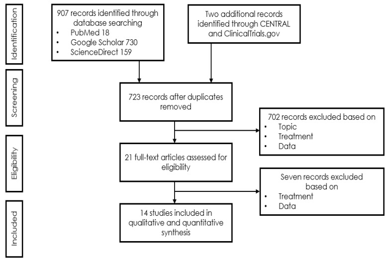
The aim of this paper is to summarise our own and to review published experience regarding the long-term outcome of intravitreal treatment for macular neovascularisation (MNV) secondary to Sorsby's fundus dystrophy (SFD). A systematic literature search using the MeSH terms [Sorsby] and [anti-vascular endothelial growth factor (VEGF)] was conducted in NCBI/PubMed, Cochrane Central Register of Controlled Trials (CENTRAL), ScienceDirect, Google Scholar and ClinicalTrials.gov to identify publications reporting anti-VEGF treatment outcomes in SFD. Treatment outcomes were extracted for this meta-analysis from 14 publications and an own patient reporting a total of 31 cases with a mean follow-up (FU) of 54 months. Both eyes were affected in ten (32.3%) instances. Heterogenous reporting limited the comparability of the outcomes. All papers in common, however, reported satisfied to excellent responses to anti-VEGF therapy if patients were diagnosed and treated immediately after onset of symptoms. Of 20 eyes, for which visual acuity was reported before and after treatment, five worsened and seven improved by more than 1 line, whereas eight eyes maintained their function by end of the follow up, and 11 eyes (55%) maintained a driving vision (Snellen VA ≥ 0.5). Of six eyes with a VA < 0.5, VA improved in one to VA ≥ 0.5, whereas of 14 eyes with an initial VA ≥ 0.5, this dropped to <0.5 despite therapy. In MNV secondary to SFD, the delay between first symptoms and access to anti-VEGF treatment determines subretinal scar formation and thereby, functional prognosis. If treated early, this is generally favourable under regular controls and a consequent anti-VEGF treatment of MNV activity.

Keywords: Sorsby; Sorsby’s fundus dystrophy; anti-VEGF treatment; choroidal neovascularisation; hereditary retinal dystrophy; long-term FU; macular neovascularization; treatment outcome.


Conflict of interest statement

The authors declare no conflict of interest. (Commercial relationships disclosures: Arthur Baston, None; Christin Gerhardt, None; Souska Zandi, None; Justus G. Garweg, AbbVie, Alcon, Chengdu Khanghong, Bayer, Novartis).

Figures

Figure 1
Figure 1
M, 35 years, M. Sorsby. Clinical image of both eyes with a significant submacular fibrovascular lesion after three courses of photodynamic therapy in the right (a) and four in the left eye (b), prior to the start of intravitreal therapy. (second panel). Same patient, fluorescein angiography (R middle (c), L early arteriovenous phase (d)) confirming a low-active predominantly classic macular neovascularisation.
Figure 2
Figure 2
Same patient, 5 years later. First reactivation of macular neovascularisation evidenced by vision loss and a small macular hemorrhage as well as newly present intraretinal fluid in OCT in the right eye (a) and macular pigment atrophy in both eyes (a,b).
Figure 3
Figure 3
Same patient, 2016, 10 years after the start of intravitreal therapy; no lesion activity after 22 intravitreal Ranibizumab injections in the right eye (a) and a remarkable progressive macular atrophy despite a stable scar in his left eye (b).
Figure 4
Figure 4
Same patient, 2021, meanwhile 51 years old. Eighteen years after diagnosis and 15 years after the start of intravitreal therapy visual acuity was maintained at Snellen 1.0 (20/20) in his right (a) and 0.16 (32/200) in his left eye (b), though reading and contrast-enhancing optical aids are required for near visual performance; no lesion activity after 22 intravitreal Ranibizumab injections in the right eye and a widely unchanged macular situation. Progressive macular scarring in both eyes. Upper panel: Clinical pictures of R + L eye (a,b), second panel, redfree picture and OCT of the right eye (c), bottom same, left eye (d). The arrows in redfree frames on the left side in Figure 4c,d indicate the location of the line scans on the right side. Note the progression of severity and extension of RPE changes, Drusen formation and choroidal sclerosis during the observation period. With consequent clinical controls and Ranibizumab treatment immediately upon first signs of lesion reactivation, his quality of life is perceived as excellent, he can follow his daily professional and private activities without relevant restrictions.
Figure 4
Figure 4
Same patient, 2021, meanwhile 51 years old. Eighteen years after diagnosis and 15 years after the start of intravitreal therapy visual acuity was maintained at Snellen 1.0 (20/20) in his right (a) and 0.16 (32/200) in his left eye (b), though reading and contrast-enhancing optical aids are required for near visual performance; no lesion activity after 22 intravitreal Ranibizumab injections in the right eye and a widely unchanged macular situation. Progressive macular scarring in both eyes. Upper panel: Clinical pictures of R + L eye (a,b), second panel, redfree picture and OCT of the right eye (c), bottom same, left eye (d). The arrows in redfree frames on the left side in Figure 4c,d indicate the location of the line scans on the right side. Note the progression of severity and extension of RPE changes, Drusen formation and choroidal sclerosis during the observation period. With consequent clinical controls and Ranibizumab treatment immediately upon first signs of lesion reactivation, his quality of life is perceived as excellent, he can follow his daily professional and private activities without relevant restrictions.
Figure 5
Figure 5
PRISMA (Preferred Reporting Items for Systematic Reviews and Meta-Analyses) search flow.

References

    1. Sorsby A., Mason M.E.J., Gardener N. A Fundus Dystrophy with Unusual Features (Late onset and dominant inheritance of a central retinal lesion showing oedema, haemorrhage and exudates developing into generalised choroidal atrophy with massive pigment proliferation) Br. J. Ophthalmol. 1949;33:67–97. doi: 10.1136/bjo.33.2.67. - DOI - PMC - PubMed
    1. Capon M.R.C., Polkinghorne P.J., Fitzke F.W., Bird A.C. Sorsby’s pseudoinflammatory macula dystrophy—Sorsby’s fundus dystrophies. Eye. 1988;2:114–122. doi: 10.1038/eye.1988.23. - DOI - PubMed
    1. Jacobson S.G., Cideciyan A.V., Regunath G., Rodriguez F.J., VanDenburgh K., Sheffield V.C., Stone E.M. Night blindness in Sorsby’s fundus dystrophy reversed by vitamin A. Nat. Genet. 1995;11:27–32. doi: 10.1038/ng0995-27. - DOI - PubMed
    1. Handsley M.M., Edwards D.R. Metalloproteinases and their inhibitors in tumor angiogenesis. Int. J. Cancer. 2005;115:849–860. doi: 10.1002/ijc.20945. - DOI - PubMed
    1. Qi J.H., Ebrahem Q., Ali M., Cutler A., Bell B., Prayson N., Sears J., Knauper V., Murphy G., Anand-Apte B. Tissue Inhibitor of Metalloproteinases-3 Peptides Inhibit Angiogenesis and Choroidal Neovascularization in Mice. PLoS ONE. 2013;8:e55667. doi: 10.1371/journal.pone.0055667. - DOI - PMC - PubMed