Review
. 2021 May 23;13(11):2557.
doi: 10.3390/cancers13112557.

Transformational Role of Medical Imaging in (Radiation) Oncology

Affiliations
Review

Transformational Role of Medical Imaging in (Radiation) Oncology

Catherine Coolens et al. Cancers (Basel). .

Abstract

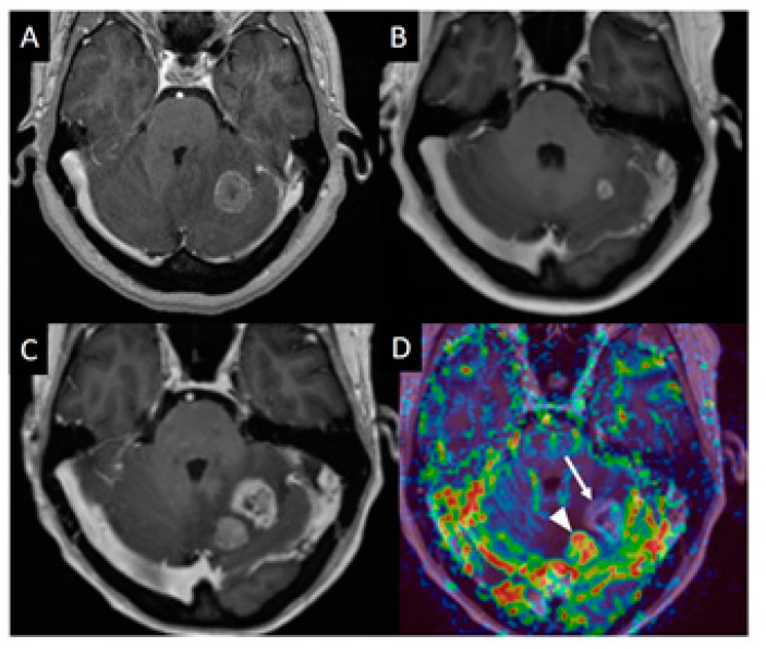
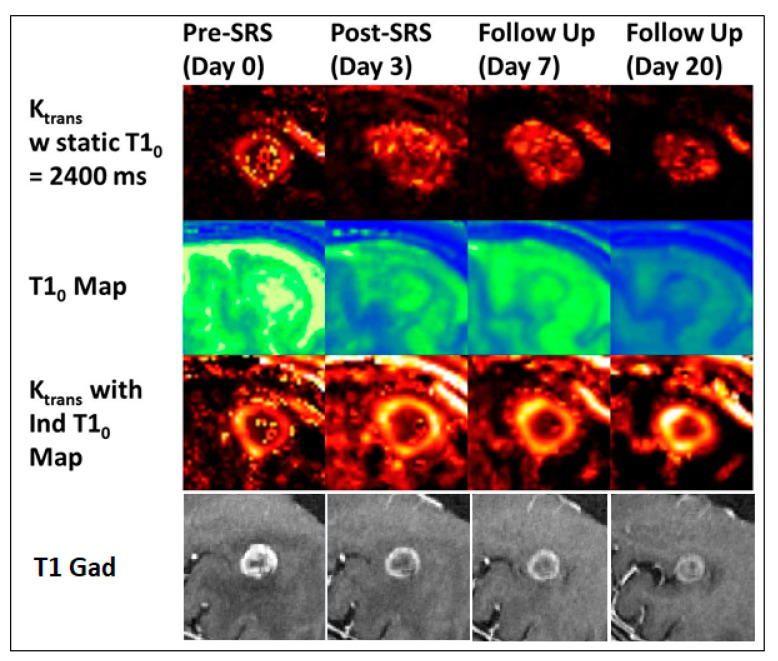
Onboard, real-time, imaging techniques, from the original megavoltage planar imaging devices, to the emerging combined MRI-Linear Accelerators, have brought a huge transformation in the ability to deliver targeted radiation therapies. Each generation of these technologies enables lethal doses of radiation to be delivered to target volumes with progressively more accuracy and thus allows shrinking of necessary geometric margins, leading to reduced toxicities. Alongside these improvements in treatment delivery, advances in medical imaging, e.g., PET, and MRI, have also allowed target volumes themselves to be better defined. The development of functional and molecular imaging is now driving a conceptually larger step transformation to both better understand the cancer target and disease to be treated, as well as how tumors respond to treatment. A biological description of the tumor microenvironment is now accepted as an essential component of how to personalize and adapt treatment. This applies not only to radiation oncology but extends widely in cancer management from surgical oncology planning and interventional radiology, to evaluation of targeted drug delivery efficacy in medical oncology/immunotherapy. Here, we will discuss the role and requirements of functional and metabolic imaging techniques in the context of brain tumors and metastases to reliably provide multi-parametric imaging biomarkers of the tumor microenvironment.

Keywords: RECIST; brain cancer; imaging biomarker; microenvironment; quantitative imaging; radiation oncology; response.


Conflict of interest statement

The authors declare no conflict of interest.

Figures

Figure 1
Figure 1
T1-weighted and DSC imaging of a left cerebellar metastasis. (A) Post contrast T1-Weighted MRI image of a 40 yo (year old) female with colon cancer and left cerebellar metastasis. (B) MRI 7 months later showed marked reduction is size after radiosurgery treatment. (C) MRI performed 11 months after treatment showed increase in size of the treated lesion and a new enhancing lesion medial to it. (D) CBV map from DSC perfusion (arrow) demonstrates mainly low perfusion. Some small foci of slightly elevated perfusion may represent foci of recurrent tumor on a background of predominant radiation necrosis.
Figure 2
Figure 2
Central section through a brain metastasis for the same patient over different imaging days showing (top) Ktrans values using a static T10 map, (middle) the voxel-based T10 map, and (bottom) Ktrans values using the individual T10 map [37].

Similar articles

Cited by

References

    1. Bujold A., Craig T., Jaffray D., Dawson L.A. Image-guided radiotherapy: Has it influenced patient outcomes? Semin. Radiat. Oncol. 2012;22:50–61. doi: 10.1016/j.semradonc.2011.09.001. - DOI - PubMed
    1. Jaffray D.A. Image-guided radiotherapy: From current concept to future perspectives. Nat. Rev. Clin. Oncol. 2012;9:688–699. doi: 10.1038/nrclinonc.2012.194. - DOI - PubMed
    1. Jaffray D.A., Chung C., Coolens C., Foltz W., Keller H., Menard C., Milosevic M., Publicover J., Yeung I. Quantitative Imaging in Radiation Oncology: An Emerging Science and Clinical Service. Semin. Radiat. Oncol. 2015;25:292–304. doi: 10.1016/j.semradonc.2015.05.002. - DOI - PubMed
    1. Hoffmann A., Oborn B., Moteabbed M., Yan S., Bortfeld T., Knopf A., Fuchs H., Georg D., Seco J., Spadea M.F., et al. MR-guided proton therapy: A review and a preview. Radiat. Oncol. 2020;15:129. doi: 10.1186/s13014-020-01571-x. - DOI - PMC - PubMed
    1. Landry G., Hua C.H. Current state and future applications of radiological image guidance for particle therapy. Med. Phys. 2018;45:e1086–e1095. doi: 10.1002/mp.12744. - DOI - PubMed

LinkOut - more resources