

Demethoxycurcumin Suppresses Human Brain Glioblastoma Multiforme GBM 8401 Cell Xenograft Tumor in Nude Mice In Vivo
- PMID: 34071132
- PMCID: PMC8197162
- DOI: 10.3390/ijms22115503
Demethoxycurcumin Suppresses Human Brain Glioblastoma Multiforme GBM 8401 Cell Xenograft Tumor in Nude Mice In Vivo
Abstract
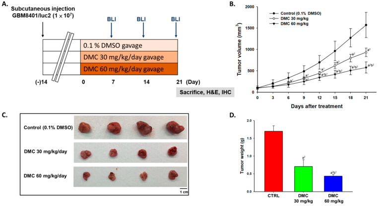
Demethoxycurcumin (DMC), a derivate of curcumin, has been shown to induce apoptotic cell death in human glioblastoma multiforme GBM 8401 cells via cell cycle arrest and induction of cell apoptosis. However, there is no report showing DMC suppresses glioblastoma multiforme cells in vivo. In the present study, we investigated the effects of DMC on GBM8401 cells in vivo. At first, we established a luciferase-expressing stable clone named GBM 8401/luc2. Second, mice were inoculated subcutaneously with GBM 8401/luc2 cells to generate a xenograft tumor mice model. After inoculation, tumor volume reached 100-120 mm3, and all mice were randomly divided into three groups: Group I was treated with 110 µL phosphate-buffered solution (PBS) containing 0.1% dimethyl sulfoxide, Group II with 30 mg/kg of DMC, and Group III with 60 mg/kg of DMC. Mice from each group were given the oral treatment of DMC by gavage for 21 days. The body weight and tumor volume were recorded every 3 days. DMC significantly decreased the tumor volumes, and 60 mg/kg treatment showed a higher decrease in tumor volumes than that of 30 mg/kg, However, DMC did not affect the body weights. The photons emitted from mice tumors were detected with Xenogen IVIS imaging system, DMC at both doses decreased the total photon flux and 60 mg/kg treatment of DMC has low total photon flux than that of 30 mg/kg. The tumor volumes and weights in 60 mg/kg treatment of DMC were lower than that of 30 mg/kg. Immunohistochemical analysis was used to measure protein expression of tumors and results showed that DMC treatment led to lightly staining with anti-Bcl-2 and -XIAP and 60 mg/kg treatment of DMC has lighter staining with anti-Bcl-2 and -XIAP than that of 30 mg/kg. The higher dose (60 mg/kg) of DMC has higher signals of cleaved-caspase-3 than that of the lower dose (30 mg/kg). Furthermore, the hematoxylin and eosin (H&E) staining of liver tissues showed no significant difference between DMC-treated and control-groups. Overall, these observations showed that DMC suppressed tumor properties in vivo and DMC may be used against human glioblastoma multiforme in the future.
Keywords: demethoxycurcumin (DMC); glioblastoma multiforme; in vivo; nude mice; xenograft tumor.
Conflict of interest statement
We wish to confirm that there are no known conflict of interest associated with this publication and there has been no significant financial support for this work that could have influenced its outcome.
Figures


Similar articles
-
Tetrandrine Suppresses Human Brain Glioblastoma GBM 8401/luc2 Cell-Xenografted Subcutaneous Tumors in Nude Mice In Vivo.Molecules. 2021 Nov 24;26(23):7105. doi: 10.3390/molecules26237105. Molecules. 2021. PMID: 34885686 Free PMC article.
-
Benzyl isothiocyanate inhibits human brain glioblastoma multiforme GBM 8401 cell xenograft tumor in nude mice in vivo.Environ Toxicol. 2018 Nov;33(11):1097-1104. doi: 10.1002/tox.22581. Epub 2018 Jul 4. Environ Toxicol. 2018. PMID: 29972272
-
Allyl Isothiocyanate (AITC) Induces Apoptotic Cell Death In Vitro and Exhibits Anti-Tumor Activity in a Human Glioblastoma GBM8401/luc2 Model.Int J Mol Sci. 2022 Sep 8;23(18):10411. doi: 10.3390/ijms231810411. Int J Mol Sci. 2022. PMID: 36142326 Free PMC article.
-
Anti-tumor Effects of Curcuminoids in Glioblastoma Multiforme: An Updated Literature Review.Curr Med Chem. 2021;28(39):8116-8138. doi: 10.2174/0929867327666201111145212. Curr Med Chem. 2021. PMID: 33176632 Review.
-
Natural bioactive molecules: An alternative approach to the treatment and control of glioblastoma multiforme.Biomed Pharmacother. 2021 Sep;141:111928. doi: 10.1016/j.biopha.2021.111928. Epub 2021 Jul 19. Biomed Pharmacother. 2021. PMID: 34323701 Review.
Cited by
-
Demethoxycurcumin induces apoptosis and reduces cell migration by affecting AKT/mTOR-dependent autophagy in human glioma U87MG and T98G cell lines.Mol Biol Rep. 2025 Sep 8;52(1):872. doi: 10.1007/s11033-025-10987-1. Mol Biol Rep. 2025. PMID: 40920279
-
Potential of Natural Products in the Treatment of Glioma: Focus on Molecular Mechanisms.Cell Biochem Biophys. 2024 Dec;82(4):3157-3208. doi: 10.1007/s12013-024-01447-x. Epub 2024 Aug 16. Cell Biochem Biophys. 2024. PMID: 39150676 Review.
References
-
- Kumar V., Aster J.C. Robbins Basic Pathology E-Book. Elsevier Health Sciences; Amsterdam, The Netherlands: 2017.
-
- Louis D.N., Perry A., Reifenberger G., von Deimling A., Figarella-Branger D., Cavenee W.K., Ohgaki H., Wiestler O.D., Kleihues P., Ellison D.W. The 2016 World Health Organization Classification of Tumors of the Central Nervous System: A summary. Acta Neuropathol. 2016;131:803–820. doi: 10.1007/s00401-016-1545-1. - DOI - PubMed
Publication types
MeSH terms
Substances
Grants and funding
LinkOut - more resources
Full Text Sources
Medical
Research Materials
