Left and Right Myocardial Functionality Assessed by Two-Dimensional Speckle-Tracking Echocardiography in Cats with Restrictive Cardiomyopathy
- PMID: 34071192
- PMCID: PMC8226601
- DOI: 10.3390/ani11061578
Left and Right Myocardial Functionality Assessed by Two-Dimensional Speckle-Tracking Echocardiography in Cats with Restrictive Cardiomyopathy
Abstract
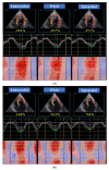
The endomyocardial form of restrictive cardiomyopathy (EMF-RCM), a primary disorder of the myocardium, is one of the diseases with poor prognosis in cats. We hypothesized that both the left and right myocardial functional abnormalities may occur in cats with EMF-RCM, causing this disease pathophysiology and clinical status. Out of the 25 animals included in this study, 10 were client-owned cats with EMF-RCM, and 15 were healthy cats. In this study, cats were assessed for layer-specific myocardial function (whole, endocardial, and epicardial) in the left ventricular longitudinal and circumferential directions, and right ventricular longitudinal direction, via two-dimensional speckle-tracking echocardiography (2D-STE). Cats with EMF-RCM had depressed left ventricular myocardial deformations both in systole (whole longitudinal strain, epicardial longitudinal strain, and endocardial circumferential strain) and diastole (early and late diastolic longitudinal strain rates, and late diastolic circumferential strain rate) compared to controls. Furthermore, some right ventricular myocardial deformations (systolic longitudinal strain in epicardial layers, and endocardial-to-epicardial strain ratio) were significantly differerent in cats with EMF-RCM. Myocardial function assessed by 2D-STE could reveal left and right myocardial dysfunction.
Keywords: endomyocardial fibrosis; feline; heart; restrictive cardiomyopathy; speckle-tracking echocardiography; strain; strain rate.
Conflict of interest statement
The authors declare no conflict of interest.
Figures


References
-
- Luis Fuentes V., Abbott J., Chetboul V., Côté E., Fox P.R., Häggström J., Kittleson M.D., Schober K., Stern J.A. ACVIM consensus statement guidelines for the classification, diagnosis, and management of cardiomyopathies in cats. J. Vet. Intern. Med. 2020;34:1062–1077. doi: 10.1111/jvim.15745. - DOI - PMC - PubMed
-
- Amaki M., Savino J., Ain D.L., Sanz J., Pedrizzetti G., Kulkarni H., Narula J., Sengupta P.P. Diagnostic concordance of echocardiography and cardiac magnetic resonance-based tissue tracking for differentiating constrictive pericarditis from restrictive cardiomyopathy. Circ. Cardiovasc. Imaging. 2014;7:819–827. doi: 10.1161/CIRCIMAGING.114.002103. - DOI - PubMed
-
- Kusunose K., Dahiya A., Popović Z.B., Motoki H., Alraies M.C., Zurick A.O., Bolen M.A., Kwon D.H., Flamm S.D., Klein A.L. Biventricular mechanics in constrictive pericarditis comparison with restrictive cardiomyopathy and impact of pericardiectomy. Circ. Cardiovasc. Imaging. 2013;6:399–406. doi: 10.1161/CIRCIMAGING.112.000078. - DOI - PubMed
Grants and funding
LinkOut - more resources
Full Text Sources
Research Materials
Miscellaneous

