

Vessel Density Loss of the Deep Peripapillary Area in Glaucoma Suspects and Its Association with Features of the Lamina Cribrosa
- PMID: 34071218
- PMCID: PMC8197842
- DOI: 10.3390/jcm10112373
Vessel Density Loss of the Deep Peripapillary Area in Glaucoma Suspects and Its Association with Features of the Lamina Cribrosa
Abstract
Purpose: To investigate the association of decreased vessel density (VD) in the deep peripapillary region and structural features of the lamina cribrosa (LC).
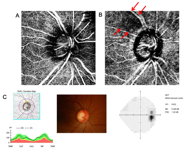
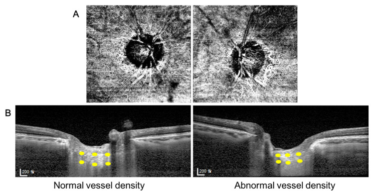
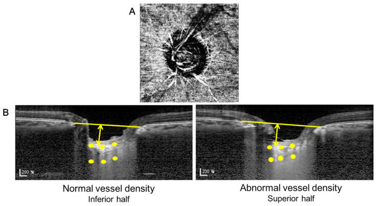
Materials and methods: 70 eyes of glaucoma suspects with enlarged cup-to-disc ratio were scanned and 51 eyes with adequate image quality were included in this study. All subjects had localized VD defects in the deep layer but intact VD in the superficial layer around the peripapillary region using optical coherence tomography angiography (OCTA). Only single-hemizone OCTA results from one eye of each subject had to fulfill the distinctive feature mentioned above to perform inter-eye and inter-hemizone comparisons. The thickness and depth of the LC, and prelaminar thickness were measured using enhanced depth imaging OCT (EDI-OCT). Paired t-tests were performed to evaluate differences in measurements of the LC and prelaminar thickness within each individual. p-values lower than 0.05 was considered to be statistically significant.
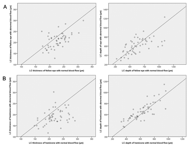
Results: Eyes with deep VD defects in the peripapillary region in OCTA had thinner LC than the fellow eyes. The hemizone with the deep VD defects in the peripapillary region had a thinner LC and a deeper depth of LC than the other hemizone in the same eye. According to logistic regression analysis, a thin LC was a significant factor associated with deep VD defect in the peripapillary region.
Conclusions: Glaucoma suspect eyes with deep VD defects in the peripapillary area exhibited structural differences in the LC. The structural changes of the LC was associated with the vessel density in the deep peripapillary layer at the stage of suspected glaucoma.
Keywords: glaucoma suspect; lamina cribrosa; optical coherence tomography angiography; peripapillary vessel density.
Conflict of interest statement
The authors report no conflict of interest. The authors alone are responsible for the content and writing of the paper.
Figures
References
-
- Chen C.-L., Bojikian K.D., Wen J.C., Zhang Q., Xin C., Mudumbai R.C., Johnstone M.A., Chen P.P., Wang R.K. Peripapillary Retinal Nerve Fiber Layer Vascular Microcirculation in Eyes with Glaucoma and Single-Hemifield Visual Field Loss. JAMA Ophthalmol. 2017;135:461–468. doi: 10.1001/jamaophthalmol.2017.0261. - DOI - PMC - PubMed
LinkOut - more resources
Full Text Sources
