. 2021 May 28;7(6):426.
doi: 10.3390/jof7060426.

A Multiomic Approach to Understand How Pleurotus eryngii Transforms Non-Woody Lignocellulosic Material

Affiliations

A Multiomic Approach to Understand How Pleurotus eryngii Transforms Non-Woody Lignocellulosic Material

Ander Peña et al. J Fungi (Basel). .

Abstract

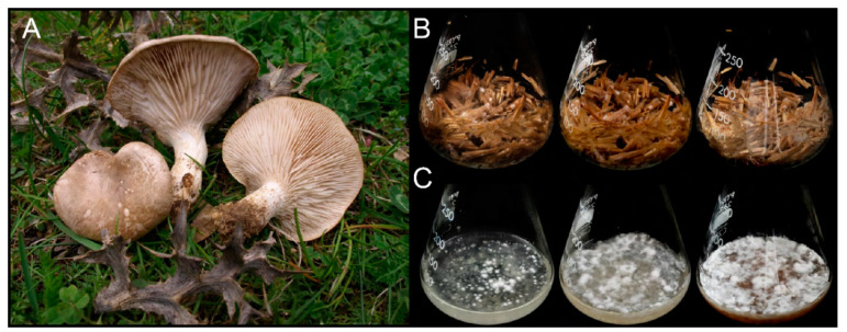
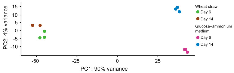
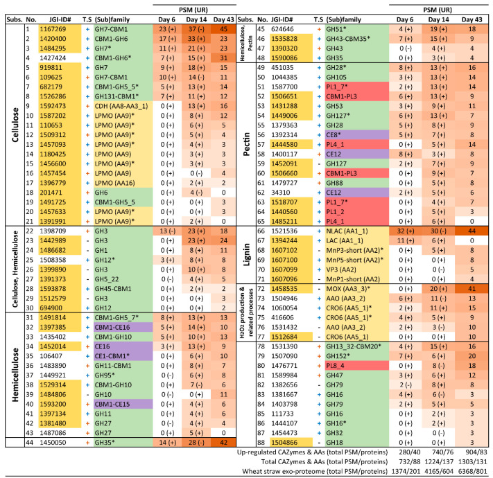
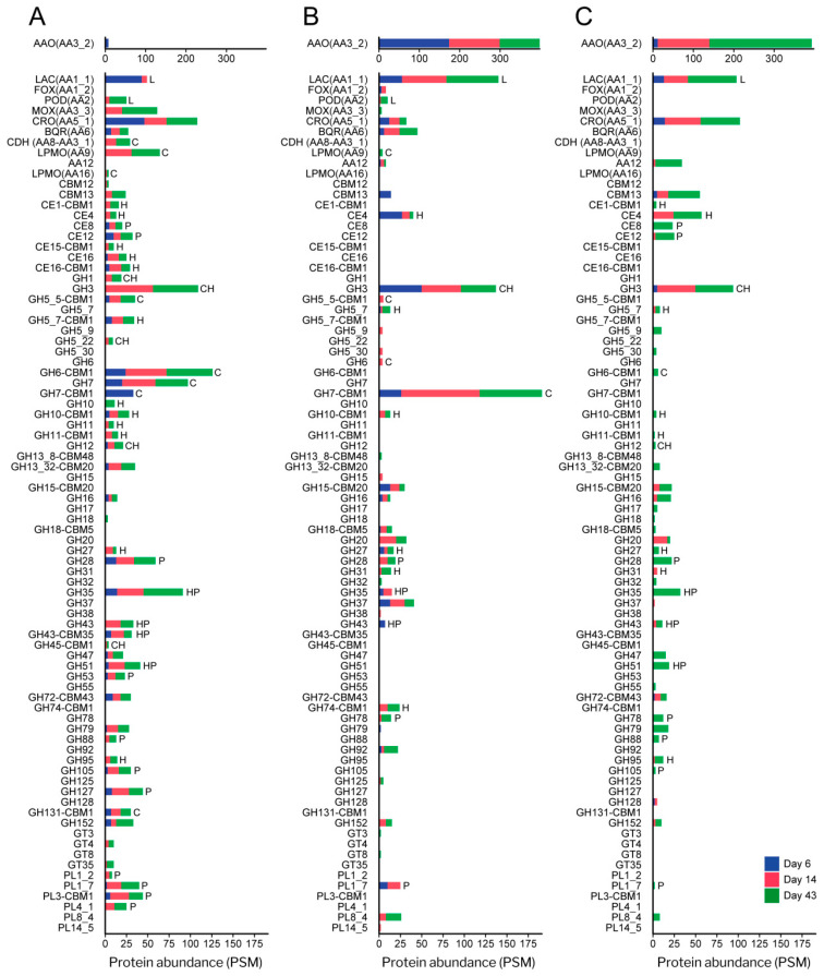
Pleurotus eryngii is a grassland-inhabiting fungus of biotechnological interest due to its ability to colonize non-woody lignocellulosic material. Genomic, transcriptomic, exoproteomic, and metabolomic analyses were combined to explain the enzymatic aspects underlaying wheat-straw transformation. Up-regulated and constitutive glycoside-hydrolases, polysaccharide-lyases, and carbohydrate-esterases active on polysaccharides, laccases active on lignin, and a surprisingly high amount of constitutive/inducible aryl-alcohol oxidases (AAOs) constituted the suite of extracellular enzymes at early fungal growth. Higher enzyme diversity and abundance characterized the longer-term growth, with an array of oxidoreductases involved in depolymerization of both cellulose and lignin, which were often up-regulated since initial growth. These oxidative enzymes included lytic polysaccharide monooxygenases (LPMOs) acting on crystalline polysaccharides, cellobiose dehydrogenase involved in LPMO activation, and ligninolytic peroxidases (mainly manganese-oxidizing peroxidases), together with highly abundant H2O2-producing AAOs. Interestingly, some of the most relevant enzymes acting on polysaccharides were appended to a cellulose-binding module. This is potentially related to the non-woody habitat of P. eryngii (in contrast to the wood habitat of many basidiomycetes). Additionally, insights into the intracellular catabolism of aromatic compounds, which is a neglected area of study in lignin degradation by basidiomycetes, were also provided. The multiomic approach reveals that although non-woody decay does not result in dramatic modifications, as revealed by detailed 2D-NMR and other analyses, it implies activation of the complete set of hydrolytic and oxidative enzymes characterizing lignocellulose-decaying basidiomycetes.

Keywords: Pleurotus eryngii; carbohydrate-active enzymes; lignin-modifying enzymes; lignocellulose transformation; metabolomics; oxidoreductases; proteomics; solid-state fermentation; transcriptomics; wheat–straw.


Conflict of interest statement

The authors declare no conflict of interest.

Figures

Figure 1
Figure 1
Pleurotus eryngii fruiting bodies (A), and monokaryotic (ATCC 90797) cultures on wheat–straw (B) and in glucose–ammonium medium (C) after 6 (left), 14 (center) and 43 (right) days of culture (image of fruit bodies courtesy of Dr J.M. Barrasa).
Figure 2
Figure 2
Principal component analysis of the log2 normalized RNA-seq read counts of 11 biological replicates of P. eryngii growing on wheat–straw and in glucose–ammonium medium for 6 and 14 days. The replicates are shown as colored circles according to the growth conditions: (i) green and brown for cultures of 6 and 14 days on wheat–straw, respectively; and purple and blue for 6 and 14 days of culture in glucose–ammonium medium, respectively.
Figure 3
Figure 3
Enrichment of up-regulated genes in P. eryngii when growing on wheat–straw compared with glucose–ammonium medium, clustered by the GO categories: CC, “Cellular Component”; BP, “Biological Process”; and MF “Molecular Function”. The bar chart shows the expected (blue) and observed (brown) number of up-regulated genes of each GO term.
Figure 4
Figure 4
Functional distribution of proteins identified in the exoproteome of P. eryngii grown on wheat–straw. (A) Venn diagram of total protein numbers (897) after 6 (blue), 14 (red) and 43 (green) days of culture. (B) Distribution into functional groups. (C) Distribution of CAZymes and oxidoreductases on 6, 14, and 43 days of culture. The number of proteins is followed by their percentage in (B,C), and their predicted functions are indicated by color codes.
Figure 5
Figure 5
Abundance in PSM values (followed by the percentage) of the main protein types, indicated by color codes, in the exoproteome on wheat–straw cultures after 6, 14, and 43 days of incubation. The central numbers correspond to the number of secreted proteins (left) and their abundance in PSM values (right). Black and yellow arcs indicate oxidoreductases and CAZymes, respectively.
Figure 6
Figure 6
Diversity of CAZymes and oxidoreductases (AA) in the exoproteome of P. eryngii grown on wheat–straw and in glucose–ammonium medium. (A) Number of up-regulated (from Figure 7) and a few only highly transcribed (from Table S5), and (B) basally expressed (from Figure 8) enzymes of each (sub)family identified in the exoproteome of wheat–straw; and (C) number of enzymes identified in glucose–ammonium medium. The length of the bars represents the total number (sum) of proteins of each CAZyme and AA (sub)family identified on day 6 (blue section), 14 (red section) and 43 (green section). Although transcriptomic data were not available for day 43, we assumed that regulation of day 14 was maintained considering that the proportion of GHs, CEs, PLs and AAs was relatively stable (Figure 4C). The substrates on which the enzymes act are indicated: C, cellulose; H, hemicellulose; L, lignin; and P, pectin. Although laccases (Lac) and PODs are the only oxidoreductases here assigned a function (acting on lignin); AAO, methanol oxidases (MOX) and CRO (producing H2O2); and benzoquinone reductases (BQR) can be considered to be indirectly involved in lignocellulose degradation.
Figure 7
Figure 7
Up-regulated CAZymes and oxidoreductases (AA) identified in the wheat–straw exoproteome. The enzymes are organized according to the substrate on which they act or the process in which they participate (column “Subs”) and then sorted from highest to lowest abundance based on total PSM values. The protein reference numbers (JGI-ID#) correspond to those in the catalog of predicted proteins from the P. eryngii ATCC 90797 genome available through JGI MycoCosm Genome Portal. Protein references over yellow background are enzymes exclusively secreted on wheat–straw. The presence (+) or absence (-) of theoretical secretion (T.S) is indicated. Symbol + in blue or orange color represents proteins predicted to be secreted by the classical secretory pathway or by the unconventional pathway, respectively. Presence (+) or absence (-) of up-regulation (UR) in wheat–straw is indicated at day 6 and 14 following the PSM values. Enzymes 78–88 have not been related to lignocellulose degradation. * Proteins found both up-regulated and highly transcribed. Colors in the (sub)family column correspond to: GH, green; oxidoreductases, yellow; PL, red; and CE, purple.
Figure 8
Figure 8
CAZymes and oxidoreductases (AA) transcribed at basal level identified in the wheat–straw exoproteome. The enzymes are organized according to the substrate on which they act or the process in which they participate (column “Subs”) and then sorted from highest to lowest abundance based on total PSM values. The protein reference numbers (JGI-ID#) correspond to those in the catalog of predicted proteins from the P. eryngii ATCC 90797 genome available through JGI MycoCosm Genome Portal. Protein references over yellow background correspond to enzymes exclusively secreted on wheat–straw. The presence (+) or absence (-) of theoretical secretion (T.S) is indicated. Symbol + in blue or orange color represents proteins predicted to be secreted by the classical secretory pathway or by the unconventional pathway, respectively. Enzymes 46–63 have not been related to lignocellulose degradation. Colors in the (sub)family column correspond to: GH, green; oxidoreductases, yellow; PL, red; CE, purple; GT (glycosyl transferase), blue; and CBM, gray.
Figure 9
Figure 9
Abundance (in PSM values) of CAZymes and oxidoreductases (AA) in the exoproteome of P. eryngii grown on wheat–straw and in glucose–ammonium medium. (A) Abundance of up-regulated (from Figure 7) and a few ones only highly transcribed (from Table S5), and (B) basally expressed (from Figure 8) enzymes of each (sub)family identified in the exoproteome on wheat–straw, and (C) abundance of enzymes identified in glucose–ammonium medium. The length of the bars represents the total abundance (sum) of proteins of each CAZyme and AA (sub)family identified on day 6 (blue section), 14 (red section) and 43 (green section). Given that transcriptomic data were not available for day 43, we assumed that regulation of day 14 was maintained also on day 43 considering that the overall composition of CAZymes and AAs was relatively stable over time regarding the proportion of GHs, CEs, PLs and AAs on wheat–straw (Figure 4C). The substrates on which the enzymes act are indicated: C, cellulose; H, hemicellulose; L, lignin; and P, pectin. Although laccases (Lac) and PODs are the only oxidoreductases here assigned a function (acting on lignin); AAO, MOX and CRO (producing H2O2); and BQR can be considered indirectly involved in lignocellulose degradation.
Figure 10
Figure 10
CBM-containing PCWDEs: (A) Catalytic domains (GHs, PLs, CEs and AA9 LPMOs) appended to CBM1 and CBM35 modules in the P. eryngii genome, transcriptome, and exoproteomes from wheat–straw, and glucose–ammonium cultures. (B,C) Abundances of catalytic domains appended and non-appended to CBM1, respectively, in the wheat–straw exoproteome after 6 (blue), 14 (red) and 43 (green) days of culture.
Figure 11
Figure 11
Abundance of main lignin-active oxidoreductases in the wheat–straw (A) and glucose–ammonium (B) exoproteomes of days 6, 14, and 43. Enzyme families: AAO, aryl–alcohol oxidases; BQR, benzoquinone reductases; CRO, copper-radical oxidases; DyP, dye-decolorizing peroxidases; LAC, laccases; MnP, manganese peroxidases; MOX, methanol oxidases; NLAC, novel laccases; VP, versatile peroxidases.
Figure 12
Figure 12
Proposed pathways for the conversion of pHBA by P. eryngii. Continuous arrows indicate enzymatic steps for which enzymes were found in the genome. The color codes indicate at least one of these enzymes expressed (from RNA-seq data) as described in the inset. Proposed enzymatic steps: (1) oxidative decarboxylase, (2) hydroxylase, (3) hydroxylation by P450s, (4) hydroxylase, (5) oxidative decarboxylase, (6) dioxygenase, (7) ring-cleaving dioxygenase, (8) 4-hydroxymuconic semialdehyde dehydrogenase, (9) maleylacetate reductase, (10) ketoacid-CoA transferase, (11) thiolase, (12) carboxylic acid reductase, (13) aldehyde dehydrogenase, (14) alcohol dehydrogenase, (15) alcohol oxidase, (16) aldehyde oxidase, (17) 4-O-methyl transferase and (18) demethylase (see Table S6 for additional information).
Figure 13
Figure 13
Degradation of wheat–straw components by P. eryngii over time. Relative abundance of: (A) major lignocellulose fractions (% of sample dry weight, with the degradation percentage with respect to day-0 in parentheses); and (B) polar aromatic compounds (vs ethylvanillin used as the internal standard at a concentration of 4 µg/g sample) in the water fraction before inoculation (Day 0, black columns) and after 6 (blue), 14 (red) and 43 (green) days of culture. Results show the average of biological triplicates and 95% confidence limits.
Figure 14
Figure 14
NMR analysis of control (uninoculated) wheat–straw and treated with P. eryngii for 43 days, acquired at the gel stage in DMSO-d6. Top. HSQC spectra displaying 13C-1H cross-correlation signals of lignin, cinnamic acid, and carbohydrate moieties. Bottom. Lignin and other aromatic structures identified including: A, β–O–4′ ether substructure (including a second S, G or T unit); A′, γ-acylated β-O-4′ ether substructure; B, phenylcoumaran substructure; C, resinol substructure; T, tricin substructure; pCA, p-coumaric acid; FA, ferulic acid; H, p-hydroxyphenyl unit, G, guaiacyl unit; S, syringyl unit; and S’, Cα-oxidized S unit (R in S’ can be a hydroxyl in carboxylic acids, a hydrogen in aldehydes, or a lignin side chain in ketones). For carbohydrates, only the anomeric signals were assigned corresponding to: Ar, arabinose units; Di, glucose moiety in disaccharide; Gl, glucose units; U, uronic acid units; X and X’, normal and acetylated xylose units; and αXr and βXr, α and β xylose reducing units.

Similar articles

Cited by

References

    1. Dahmen N., Lewandowski I., Zibek S., Weidtmann A. Integrated lignocellulosic value chains in a growing bioeconomy: Status quo and perspectives. GCB Bioenergy. 2019;11:107–117. doi: 10.1111/gcbb.12586. - DOI
    1. Himmel M.E., Ding S.Y., Johnson D.K., Adney W.S., Nimlos M.R., Brady J.W., Foust T.D. Biomass recalcitrance: Engineering plants and enzymes for biofuels production. Science. 2007;315:804–807. doi: 10.1126/science.1137016. - DOI - PubMed
    1. Ragauskas A.J., Williams C.K., Davison B.H., Britovsek G., Cairney J., Eckert C.A., Frederick W.J., Hallett J.P., Leak D.J., Liotta C.L., et al. The path forward for biofuels and biomaterials. Science. 2006;311:484–489. doi: 10.1126/science.1114736. - DOI - PubMed
    1. Schutyser W., Renders T., Van den Bosch S., Koelewijn S.F., Beckham G.T., Sels B.F. Chemicals from lignin: An interplay of lignocellulose fractionation, depolymerisation, and upgrading. Chem. Soc. Rev. 2018;47:852–908. doi: 10.1039/C7CS00566K. - DOI - PubMed
    1. Kobayashi A., Fukuoka A. Synthesis and utilisation of sugar compounds derived from lignocellulosic biomass. Green Chem. 2013;15:1740–1763. doi: 10.1039/c3gc00060e. - DOI