Challenging Safety and Efficacy of Retinal Gene Therapies by Retinogenesis
- PMID: 34071252
- PMCID: PMC8198227
- DOI: 10.3390/ijms22115767
Challenging Safety and Efficacy of Retinal Gene Therapies by Retinogenesis
Abstract
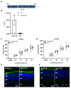
Gene-expression programs modulated by transcription factors (TFs) mediate key developmental events. Here, we show that the synthetic transcriptional repressor (TR; ZF6-DB), designed to treat Rhodopsin-mediated autosomal dominant retinitis pigmentosa (RHO-adRP), does not perturb murine retinal development, while maintaining its ability to block Rho expression transcriptionally. To express ZF6-DB into the developing retina, we pursued two approaches, (i) the retinal delivery (somatic expression) of ZF6-DB by Adeno-associated virus (AAV) vector (AAV-ZF6-DB) gene transfer during retinogenesis and (ii) the generation of a transgenic mouse (germ-line transmission, TR-ZF6-DB). Somatic and transgenic expression of ZF6-DB during retinogenesis does not affect retinal function of wild-type mice. The P347S mouse model of RHO-adRP, subretinally injected with AAV-ZF6-DB, or crossed with TR-ZF6-DB or shows retinal morphological and functional recovery. We propose the use of developmental transitions as an effective mode to challenge the safety of retinal gene therapies operating at genome, transcriptional, and transcript levels.
Keywords: adeno-associated virus (AAV); autosomal dominant; gene therapy; retinal degeneration; retinitis pigmentosa; rhodopsin; transcription; zinc finger.
Conflict of interest statement
The authors declare no conflict of interest.
Figures


Similar articles
-
AAV delivery of wild-type rhodopsin preserves retinal function in a mouse model of autosomal dominant retinitis pigmentosa.Hum Gene Ther. 2011 May;22(5):567-75. doi: 10.1089/hum.2010.140. Epub 2011 Mar 7. Hum Gene Ther. 2011. PMID: 21126223 Free PMC article.
-
Effect of AAV-Mediated Rhodopsin Gene Augmentation on Retinal Degeneration Caused by the Dominant P23H Rhodopsin Mutation in a Knock-In Murine Model.Hum Gene Ther. 2020 Jul;31(13-14):730-742. doi: 10.1089/hum.2020.008. Epub 2020 Jun 26. Hum Gene Ther. 2020. PMID: 32394751
-
Long-term rescue of retinal structure and function by rhodopsin RNA replacement with a single adeno-associated viral vector in P23H RHO transgenic mice.Hum Gene Ther. 2012 Apr;23(4):356-66. doi: 10.1089/hum.2011.213. Epub 2012 Mar 28. Hum Gene Ther. 2012. PMID: 22289036 Free PMC article.
-
Therapy in Rhodopsin-Mediated Autosomal Dominant Retinitis Pigmentosa.Mol Ther. 2020 Oct 7;28(10):2139-2149. doi: 10.1016/j.ymthe.2020.08.012. Epub 2020 Aug 25. Mol Ther. 2020. PMID: 32882181 Free PMC article. Review.
-
Gene augmentation for adRP mutations in RHO.Cold Spring Harb Perspect Med. 2014 Jul 18;4(9):a017400. doi: 10.1101/cshperspect.a017400. Cold Spring Harb Perspect Med. 2014. PMID: 25037104 Free PMC article. Review.
Cited by
-
Gene regulatory and gene editing tools and their applications for retinal diseases and neuroprotection: From proof-of-concept to clinical trial.Front Neurosci. 2022 Oct 20;16:924917. doi: 10.3389/fnins.2022.924917. eCollection 2022. Front Neurosci. 2022. PMID: 36340792 Free PMC article. Review.
-
Retinitis Pigmentosa: Novel Therapeutic Targets and Drug Development.Pharmaceutics. 2023 Feb 17;15(2):685. doi: 10.3390/pharmaceutics15020685. Pharmaceutics. 2023. PMID: 36840007 Free PMC article. Review.
-
Gene Therapy for Rhodopsin Mutations.Cold Spring Harb Perspect Med. 2022 Aug 8;12(9):a041283. doi: 10.1101/cshperspect.a041283. Online ahead of print. Cold Spring Harb Perspect Med. 2022. PMID: 35940643 Free PMC article.
-
A DNA base-specific sequence interposed between CRX and NRL contributes to RHODOPSIN expression.Sci Rep. 2024 Nov 1;14(1):26313. doi: 10.1038/s41598-024-76664-8. Sci Rep. 2024. PMID: 39487168 Free PMC article.
-
miR-181a/b downregulation: a mutation-independent therapeutic approach for inherited retinal diseases.EMBO Mol Med. 2022 Nov 8;14(11):e15941. doi: 10.15252/emmm.202215941. Epub 2022 Oct 4. EMBO Mol Med. 2022. PMID: 36194668 Free PMC article.
References
-
- Russell S., Bennett J., Wellman J.A., Chung D.C., Yu Z.F., Tillman A., Wittes J., Pappas J., Elci O., McCague S., et al. Efficacy and safety of voretigene neparvovec (AAV2-hRPE65v2) in patients with RPE65-mediated inherited retinal dystrophy: A randomised, controlled, open-label, phase 3 trial. Lancet. 2017;390:849–860. doi: 10.1016/S0140-6736(17)31868-8. - DOI - PMC - PubMed
MeSH terms
Substances
LinkOut - more resources
Full Text Sources
Medical
Molecular Biology Databases
Research Materials
Miscellaneous

