. 2021 May 28;11(6):800.
doi: 10.3390/biom11060800.

Disulfide and Fully Reduced HMGB1 Induce Different Macrophage Polarization and Migration Patterns

Affiliations

Disulfide and Fully Reduced HMGB1 Induce Different Macrophage Polarization and Migration Patterns

Henna Salo et al. Biomolecules. .

Abstract

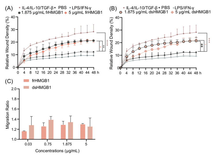
Macrophage plasticity enables cells to obtain different functions over a broad proinflammatory and repairing spectrum. In different conditions, macrophages can be induced by high-mobility group box 1 (HMGB1), a nuclear DNA-binding protein that activates innate immunity, to polarize towards a pro- (M1) or anti-inflammatory (M2) phenotype. In this study, we investigated the phenotypes of murine bone-marrow-derived macrophages (BMDMs) induced by different HMGB1 redox isoforms in depth. Our results demonstrate that disulfide HMGB1 (dsHMGB1) induces a unique macrophage phenotype that secretes pro-inflammatory cytokines, rather than inducing metabolic changes leading to nitric oxide production. Fully reduced HMGB1 (frHMGB1) did not induce macrophage polarization. The migrating function of BMDMs was measured by scratch assay after the stimulation with dsHMGB1 and frHMGB1. Both dsHMGB1 and frHMGB1 induced cell migration. We found that dsHMGB1 mediates cytokine secretion and cellular motility, mainly through toll-like receptor 4 (TLR4). Importantly, our data shows that dsHMGB1 and frHMGB1 induce distinct BMDM polarization phenotypes, and that dsHMGB1 induces a unique phenotype differing from the classical proinflammatory macrophage phenotype.

Keywords: HMGB1; RAGE; TLR4; dsHMGB1; frHMGB1; inflammation; macrophages; migration; polarization.


Conflict of interest statement

The authors declare no conflict of interest.

Figures

Figure 1
Figure 1
LPS/IFN-γ induce M1 polarization, while IL-4/IL-10/TGF-β induce M2 polarization. (A) IL-6, TNF-α, and nitrite secretion from BMDM supernatants after 24 h stimulation with LPS/IFN-γ or IL-4/IL-10/TGF-β, and (B) Arg1 gene expression in murine BMDMs stimulated with LPS/IFN-γ or IL-4/IL-10/TGF-β. (C,D) The cell migration ability of BMDMs stimulated with LPS/IFN-γ and IL-4/IL-10/TGF-β was measured using a scratch assay. (C) Representative images of scratch assay, and (D) the relative wound density of BMDMs stimulated with LPS/IFN-γ and IL-4/IL-10/TGF-β. Data represent n = 3 (mean + SD). Statistical analysis was performed using (A) a one-sample t-test, (B) t-test, and (D) a one-way ANOVA. * p ≤ 0.05, ** p ≤ 0.01, *** p ≤ 0.001, **** p < 0.0001.
Figure 2
Figure 2
dsHMGB1 induces cytokine secretion but not nitrite production. (A) IL-6, (B) IL-10, (C) TNF-α, and (D) nitrite production by BMDMs in response to dsHMGB1 and frHMGB1 after 24 h incubation. Data represent n = 3–4 (mean + SD). Statistical analysis was performed using a one-sample t-test. * p ≤ 0.05, ** p ≤ 0.01, *** p ≤ 0.001.
Figure 3
Figure 3
dsHMGB1 induces the upregulation of M1 signature genes, but in a different kinetic pattern to LPS/IFN-γ stimulation. Gene expression of (A) Il6, (B) Tnf, (C) Nos2, (D) Arg1, and (E) Il10 in response to 5 μg/mL dsHMGB1 at different time points. Groups treated with PBS, LPS/IFN-γ, and IL-4/IL-10/TGF-β were included for comparison. Data represent n = 3. Statistical comparisons were made using a Brown–Forsythe and a Welch ANOVA with Dunnett’s T3 multiple comparisons test. * represents comparisons for the same treatment between different time points; # represents comparisons between treatments comparing the same time point. */# p ≤ 0.05, **/## p ≤ 0.01. The p-values of dsHMGB1 against IL-4/IL-10/TGF-β stimulation are presented in Supplementary Table S1.
Figure 4
Figure 4
frHMGB1 induces a gene expression pattern that differs from classical M1 and M2 cells. BMDM gene expression of (A) Il6, (B) Tnf, (C) Nos2, (D) Arg1, and (E) Il10 in response to 5 μg/mL frHMGB1 at different time points. Groups treated with PBS, LPS/IFN-γ, and IL-4/IL-10/TGF-β were included for comparison. Data represent n = 3. Statistical comparisons were performed using a Brown–Forsythe and a Welch ANOVA with Dunnett’s T3 multiple comparisons test. * represents comparisons for the same treatment between different time points; # represents comparisons between treatments comparing the same time point. */# p ≤ 0.05, ## p ≤ 0.01, ### p ≤ 0.001, #### p < 0.0001.
Figure 5
Figure 5
frHMGB1 and dsHMGB1 induce BMDM mobility. Confluent BMDMs were scratched and incubated with PBS, LPS/IFN-γ, IL-4/IL-10/TGF-β, frHMGB1, or dsHMGB1. The mobility changes of BMDMs after adding frHMGB1 (1.875 μg/mL or 5 μg/mL) or dsHMGB1 (1.875 μg/mL or 5 μg/mL) are shown in (A,B), respectively. (C) The migration ratio for BMDMs incubated with frHMGB1 or dsHMGB1 at different concentrations after 48 h. Data represent n = 3–4 (mean + SD). Statistical comparisons were performed using a two-way ANOVA on the area under the curve (A,B) or a two-way ANOVA with Sidak’s multiple comparisons. * p ≤ 0.05, ** p ≤ 0.01, *** p ≤ 0.001, **** p < 0.0001.
Figure 6
Figure 6
The expression of Cxcr4, Tlr4, and Ager in BMDMs is downregulated after dsHMGB1 and frHMGB1 stimulation. (A) Cxcr4, Tlr4, and Ager expression in unstimulated BMDMs. Data represent Ct values n = 3 (interquartile range). The gene expression of (B) Cxcr4, (C) Tlr4, and (D) Ager was determined from BMDMs incubated with LPS/IFN-γ, IL-4/IL-10/TGF-β, 5 μg/mL frHMGB1, or 5 μg/mL dsHMGB1 for 24 h. Data represent n = 3 (mean ± SD). Statistical comparisons were performed using an ANOVA and a Dunnett’s multiple comparisons test. ns: not significant; * p ≤ 0.05, ** p ≤ 0.01, *** p ≤ 0.001.
Figure 7
Figure 7
dsHMGB1 induces migration in RAGE KO BMDMs. Migration ratio for a dose titration with (A) frHMGB1 and (B) dsHMGB1 in RAGE KO and WT BMDMs. Gene expression of (C) Il6, (D) Nos2, and (E) Arg1 in WT and RAGE KO BMDMs treated with PBS (negative control), LPS/IFN-γ, IL-4/IL-10/TGF-β, 5 μg/mL frHMGB1, or 5 μg/mL dsHMGB1 for 4 h. Data represent n = 3–4 (mean + SD). Statistical analysis was performed using (A) a two-way ANOVA with Sidak’s multiple comparisons or (CE) a Welch’s t-test. * p ≤ 0.05, *** p ≤ 0.001.
Figure 8
Figure 8
TLR4 inhibition reduces dsHMGB1-induced migration. Migration ratio data for (A) WT and (B) RAGE KO BMDMs treated with 1.875 μg/mL frHMGB1 or 1.875 μg/mL dsHMGB1 for 48 h in the presence or absence of 5 μM CLI095, a TLR4 inhibitor. Data represent n = 4 mice (mean + SD). Statistical comparisons were performed using a two-way ANOVA and Sidak’s multiple comparisons test. * p ≤ 0.05, ** p ≤ 0.01, *** p ≤ 0.001, **** p < 0.0001.

References

    1. Wang H., Bloom O., Zhang M., Vishnubhakat J.M., Ombrellino M., Che J., Frazier A., Yang H., Ivanova S., Borovikova L., et al. HMG-1 as a late mediator of endotoxin lethality in mice. Science. 1999;285:248–251. doi: 10.1126/science.285.5425.248. - DOI - PubMed
    1. Andersson U., Yang H., Harris H. High-mobility group box 1 protein (HMGB1) operates as an alarmin outside as well as inside cells. Semin. Immunol. 2018;38:40–48. doi: 10.1016/j.smim.2018.02.011. - DOI - PubMed
    1. Ferrara M., Chialli G., Ferreira L.M., Ruggieri E., Careccia G., Preti A., Piccirillo R., Bianchi M.E., Sitia G., Venereau E. Oxidation of HMGB1 Is a Dynamically Regulated Process in Physiological and Pathological Conditions. Front. Immunol. 2020;11:1122. doi: 10.3389/fimmu.2020.01122. - DOI - PMC - PubMed
    1. Bonaldi T., Talamo F., Scaffidi P., Ferrera D., Porto A., Bachi A., Rubartelli A., Agresti A., Bianchi M.E. Monocytic cells hyperacetylate chromatin protein HMGB1 to redirect it towards secretion. EMBO J. 2003;22:5551–5560. doi: 10.1093/emboj/cdg516. - DOI - PMC - PubMed
    1. Xue J., Suarez J.S., Minaai M., Li S., Gaudino G., Pass H.I., Carbone M., Yang H. HMGB1 as a therapeutic target in disease. J. Cell. Physiol. 2021;236:3406–3419. doi: 10.1002/jcp.30125. - DOI - PMC - PubMed

Publication types

LinkOut - more resources