

The Emerging Role of HDACs: Pathology and Therapeutic Targets in Diabetes Mellitus
- PMID: 34071497
- PMCID: PMC8228721
- DOI: 10.3390/cells10061340
The Emerging Role of HDACs: Pathology and Therapeutic Targets in Diabetes Mellitus
Abstract
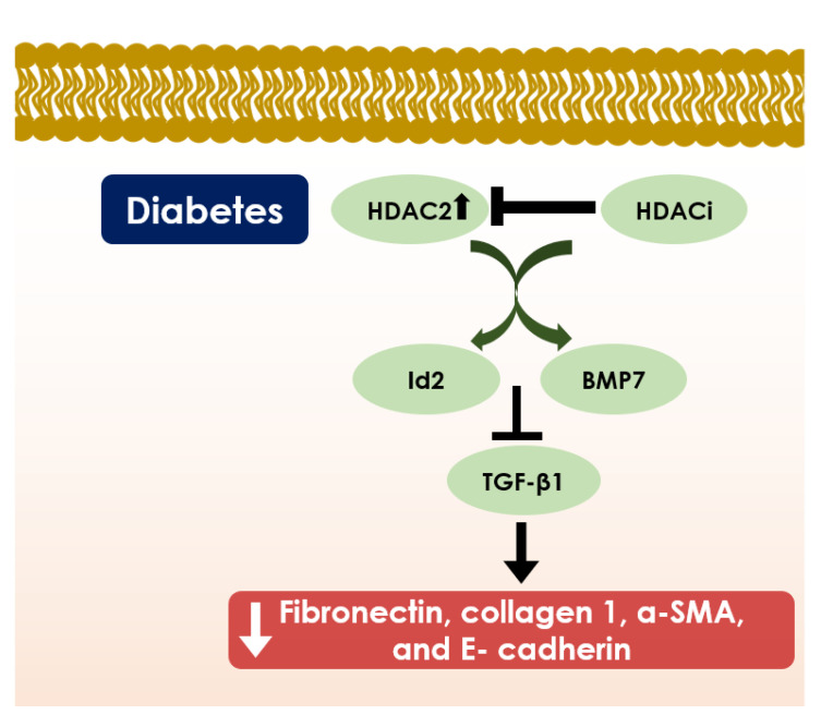
Diabetes mellitus (DM) is one of the principal manifestations of metabolic syndrome and its prevalence with modern lifestyle is increasing incessantly. Chronic hyperglycemia can induce several vascular complications that were referred to be the major cause of morbidity and mortality in DM. Although several therapeutic targets have been identified and accessed clinically, the imminent risk of DM and its prevalence are still ascending. Substantial pieces of evidence revealed that histone deacetylase (HDAC) isoforms can regulate various molecular activities in DM via epigenetic and post-translational regulation of several transcription factors. To date, 18 HDAC isoforms have been identified in mammals that were categorized into four different classes. Classes I, II, and IV are regarded as classical HDACs, which operate through a Zn-based mechanism. In contrast, class III HDACs or Sirtuins depend on nicotinamide adenine dinucleotide (NAD+) for their molecular activity. Functionally, most of the HDAC isoforms can regulate β cell fate, insulin release, insulin expression and signaling, and glucose metabolism. Moreover, the roles of HDAC members have been implicated in the regulation of oxidative stress, inflammation, apoptosis, fibrosis, and other pathological events, which substantially contribute to diabetes-related vascular dysfunctions. Therefore, HDACs could serve as the potential therapeutic target in DM towards developing novel intervention strategies. This review sheds light on the emerging role of HDACs/isoforms in diabetic pathophysiology and emphasized the scope of their targeting in DM for constituting novel interventional strategies for metabolic disorders/complications.
Keywords: HDACi; HDACs; diabetes mellitus; glucose metabolism; histone deacetylase; histone deacetylase inhibitor; insulin release; sirtuin activaton; sirtuins.
Conflict of interest statement
The authors declare no conflict of interest.
Figures
Similar articles
-
Histone deacetylases and inhibitors in diabetes mellitus and its complications.Biomed Pharmacother. 2024 Aug;177:117010. doi: 10.1016/j.biopha.2024.117010. Epub 2024 Jun 27. Biomed Pharmacother. 2024. PMID: 38941890 Review.
-
Zinc-dependent histone deacetylases: Potential therapeutic targets for arterial hypertension.Biochem Pharmacol. 2022 Aug;202:115111. doi: 10.1016/j.bcp.2022.115111. Epub 2022 May 28. Biochem Pharmacol. 2022. PMID: 35640713 Review.
-
Exploring histone deacetylases in type 2 diabetes mellitus: pathophysiological insights and therapeutic avenues.Clin Epigenetics. 2024 Jun 11;16(1):78. doi: 10.1186/s13148-024-01692-0. Clin Epigenetics. 2024. PMID: 38862980 Free PMC article. Review.
-
HDAC4: an emerging target in diabetes mellitus and diabetic complications.Eur J Med Res. 2025 May 30;30(1):429. doi: 10.1186/s40001-025-02697-y. Eur J Med Res. 2025. PMID: 40448151 Free PMC article. Review.
-
Non-sirtuin histone deacetylases in the control of cardiac aging.J Mol Cell Cardiol. 2015 Jun;83:14-20. doi: 10.1016/j.yjmcc.2015.03.010. Epub 2015 Mar 16. J Mol Cell Cardiol. 2015. PMID: 25791169 Free PMC article. Review.
Cited by
-
Mechanisms of Insulin Signaling as a Potential Therapeutic Method in Intestinal Diseases.Cells. 2024 Nov 14;13(22):1879. doi: 10.3390/cells13221879. Cells. 2024. PMID: 39594627 Free PMC article. Review.
-
β-Hydroxybutyrate upregulates FGF21 expression through inhibition of histone deacetylases in hepatocytes.Open Life Sci. 2022 Aug 10;17(1):856-864. doi: 10.1515/biol-2022-0095. eCollection 2022. Open Life Sci. 2022. PMID: 36045720 Free PMC article.
-
Multifunctional nanoparticle-mediated combining therapy for human diseases.Signal Transduct Target Ther. 2024 Jan 1;9(1):1. doi: 10.1038/s41392-023-01668-1. Signal Transduct Target Ther. 2024. PMID: 38161204 Free PMC article. Review.
-
Emerging Neuroprotective Strategies: Unraveling the Potential of HDAC Inhibitors in Traumatic Brain Injury Management.Curr Neuropharmacol. 2024;22(14):2298-2313. doi: 10.2174/1570159X22666240128002056. Curr Neuropharmacol. 2024. PMID: 38288835 Free PMC article. Review.
-
Latest in Cellular Pathology Research.Cells. 2022 Dec 14;11(24):4037. doi: 10.3390/cells11244037. Cells. 2022. PMID: 36552801 Free PMC article.
References
-
- Rivera-Mancía S., Trujillo J., Chaverri J.P. Utility of curcumin for the treatment of diabetes mellitus: Evidence from pre-clinical and clinical studies. J. Nutr. Intermed. Metab. 2018;14:29–41. doi: 10.1016/j.jnim.2018.05.001. - DOI
Publication types
MeSH terms
Substances
LinkOut - more resources
Full Text Sources
Medical
