. 2021 May 28;13(11):2673.
doi: 10.3390/cancers13112673.

Phase I Trial of Prophylactic Donor-Derived IL-2-Activated NK Cell Infusion after Allogeneic Hematopoietic Stem Cell Transplantation from a Matched Sibling Donor

Affiliations

Phase I Trial of Prophylactic Donor-Derived IL-2-Activated NK Cell Infusion after Allogeneic Hematopoietic Stem Cell Transplantation from a Matched Sibling Donor

Raynier Devillier et al. Cancers (Basel). .

Abstract

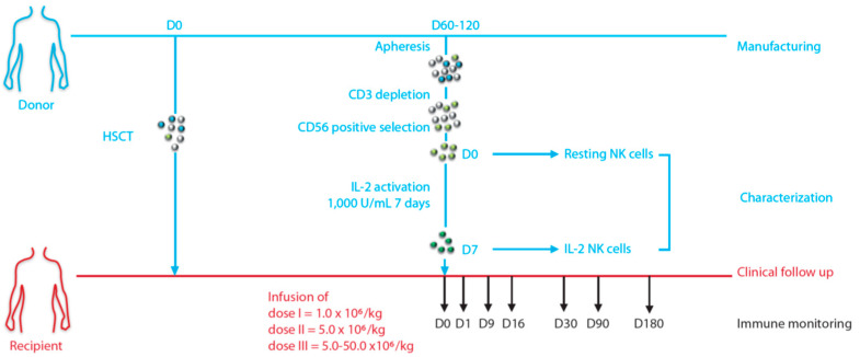
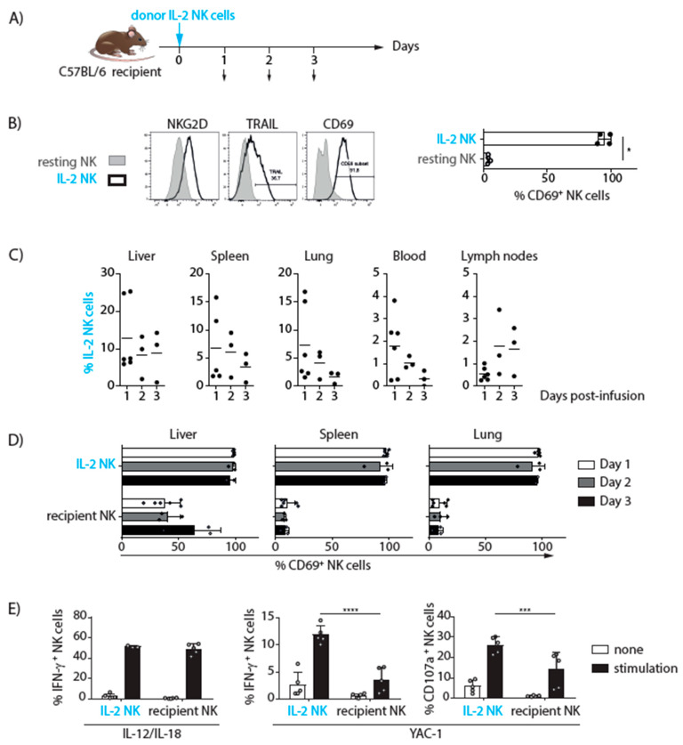
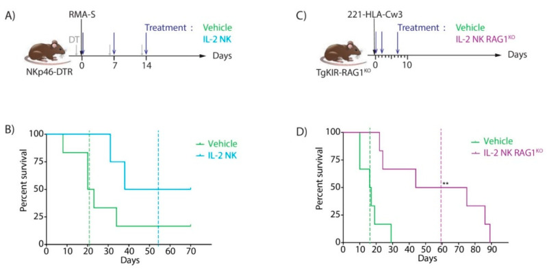
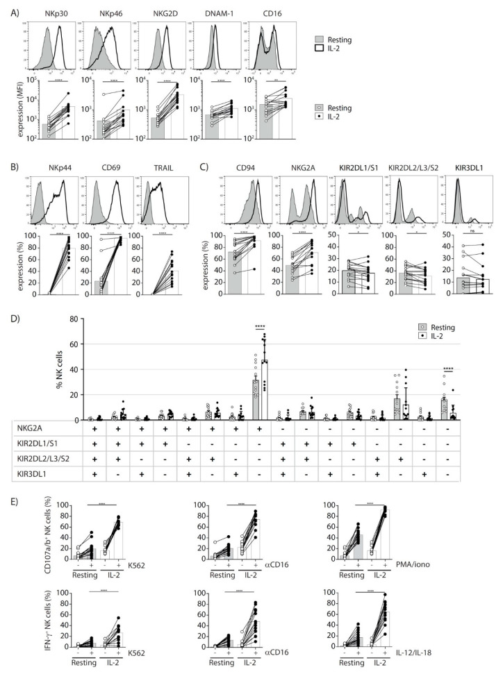
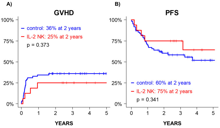
Background: NK cell-based immunotherapy to prevent relapse after allogeneic transplantation is an appealing strategy because NK cells can provide strong antitumor effect without inducing graft-versus-host disease (GVHD). Thus, we designed a phase-I clinical trial evaluating the safety of a prophylactic donor-derived ex vivo IL-2 activated NK cell (IL-2 NK) infusion after allo-HSCT for patients with hematologic malignancies. Methods: Donor NK cells were purified and cultured ex vivo with IL-2 before infusion, at three dose levels. To identify the maximum tolerated dose was the main objective. In addition, we performed phenotypical and functional characterization of the NK cell therapy product, and longitudinal immune monitoring of NK cell phenotype in patients. Results: Compared to unstimulated NK cells, IL-2 NK cells expressed higher levels of activating receptors and exhibited increased degranulation and cytokine production in vitro. We treated 16 patients without observing any dose-limiting toxicity. At the last follow up, 11 out of 16 treated patients were alive in complete remission of hematologic malignancies without GVHD features and immunosuppressive treatment. Conclusions: Prophylactic donor-derived IL-2 NK cells after allo-HSCT is safe with low incidence of GVHD. Promising survivals and IL-2 NK cell activated phenotype may support a potential clinical efficacy of this strategy.

Keywords: IL-2-activated NK cells; allogeneic hematopoietic stem cell transplantation; antitumor immunity; cellular immunotherapy.


Conflict of interest statement

E.V. is an employee and shareholder of Innate Pharma. S.U. is shareholder of Innate Pharma. The other authors declare no conflict of interest.

Figures

Figure 1
Figure 1
Design of the clinical trial: DLI-NK NCT01853358.
Figure 2
Figure 2
Monitoring of donor IL-2 NK cell phenotype and function in a mouse model. (A) Experimental scheme: donor IL-2 NK cells expressing the marker CD45.1+ were injected into congenic CD45.2+ recipient mice and their distribution, phenotype, and functional activity were monitored on day 0 before infusion and days 1, 2, and 3 post-infusion by flow cytometry. (B) Representative flow cytometry histograms showing the expression of NKG2D, TRAIL, and CD69 in resting and IL-2 NK cells before infusion (left panel). Percentages of CD69 expression in resting and IL-2 NK cells before infusion (right panel). Statistical analysis was performed with a Mann–Whitney test. * p < 0.05. (C) Percentages of donor IL-2 NK cells out of total NK cell populations in liver, spleen, lung, blood, and lymph nodes at the indicated post-infusion time points. (D) Percentages of CD69+ cells in donor (CD45.1+) and recipient (CD45.2+) NK cells in liver, spleen, and lung at days 1, 2, and 3 post-infusion. (C-D) Data were pooled from 3 experiments including one experiment with 3 mice analyzed at day 1, one experiment with 1 mouse analyzed at each time point and one experiment with 2 mice analyzed at each time point. Statistical analysis was performed with a one-way (C) or two-way ANOVA test (D). (E) Splenocytes from recipient mice were harvested 24 h post-infusion and activated ex vivo with or without a mix of IL-12 and IL-18 or the YAC-1 tumor cell line. The percentages of IFN-γ+ and CD107a+ donor (CD45.1+) and recipient (CD45.2+) NK cells are shown. Data were pooled from 2 experiments including one experiment with 1 mouse analyzed at each time point and one experiment with 2 mice analyzed at each time point. Statistical analysis was performed with a two-way ANOVA and Sidak’s multiple comparaison test. *** p = 0.0004 and **** p < 0.0001.
Figure 3
Figure 3
Anti-tumor effect of donor IL-2 NK cells in mouse models. (A) Experimental scheme: NKp46-DTR mice were treated by diphtheria toxin (DT) 1 day before and on days 6 and 13 after injection of the NK cell-sensitive tumor cell line RMA-S (day 0). Donor IL-2 NK cells or vehicle only were infused on days 0, 7, and 14. (B) Survival rates after RMA-S injection in NK cell-deficient mice treated with IL-2 NK cells (blue line) or vehicle only (green line). Median survival of IL-2 NK-treated (dotted blue line) and control group (dotted green line). (C) Experimental scheme: 221/HLA-Cw3 human tumor cells were injected in TgKIR-RAG1KO mice on day 0 and treated with either vehicle only or donor IL-2 NK cells from RAG1KO mice on days 0, 2, and 7. (D) Survival rate of tumor-bearing TgKIR-RAG1KO mice upon treatment with either PBS (green line) or IL-2 NK cells (purple line). Median survival for each group is represented by dotted lines. Log rank test, ** p = 0.0042.
Figure 4
Figure 4
Phenotypic and functional characterization of resting and IL-2-activated donor NK cells. (AC) Representative flow cytometry histograms (upper panels) and expression levels for each donor (mean fluorescence intensity, MFI, or % as indicated, lower panels), illustrating (A) NKp30, NKp46, NKG2D, DNAM-1, and CD16; (B) NKp44, CD69, and TRAIL; (C) CD94, NKG2A, KIR2DL1/S1, KIR2DL2/L3/S2, and KIR3DL1 expression before (resting) or after IL-2 activation. (D) The percentages of NK cells expressing the indicated markers for resting (grey histograms) and IL-2 activated NK cells (white histograms). (E) Functional analysis of resting versus IL-2-activated NK cells from the same donors. NK cells were stimulated or not during 4 h with the tumor cell line K562 (left panels), anti-CD16 antibodies (middle panels), and PMA and ionomycin or IL-12 and IL-18 (right panels). CD107a/b expression (upper panels) and IFN-γ production (lower panels) are shown for each donor. Statistical analysis was performed with a Wilcoxon test, *p = 0.0110, **p = 0.0013, **** p < 0.0001; ns, no significant (AC), a two-way ANOVA test and Sidak’s multiple comparaison test, **** p < 0.0001, only significant p values are indicated (D) and a one-way ANOVA test, only the p values comparing the responsiveness of resting versus IL-2 activated NK cells are indicated, **** p < 0.0001 (E).
Figure 5
Figure 5
Outcome after IL-2 NK cell infusion. GVHD (A) and PFS (B) of the cohort of patients treated with donor IL-2 NK cells (patients in the clinical trial, blue line) vs. historical control cohort (red line).
Figure 6
Figure 6
Immune monitoring by FACS after IL-2 NK infusion. (A) B, T, γδ-T, and NK cell frequencies between day+0 and day+1 after IL-2 NK cell infusion (n = 12). (B) NK cell functional assays before (day+0) and after (day+9) IL-2 NK infusion: CD107a, IFN-γ, TNF-α, and MIP-1α expression on NK cells after culturing PBMCs with or w/o K562 at an E:T ratio of 10:1 for 4 h (n = 9). (C) Longitudinal immune monitoring of the NK cell maturation markers NKG2A and CD57 (n = 9). (D) Longitudinal analyses of CD107a, IFN-γ, TNF-α, and MIP-1β expression on NK cells. PBMCs were co-cultured for 4 h with or without (not shown) K562 at an E:T of 10:1 (n = 7). Statistical tests were paired Wilcoxon (A and B) and paired Kruskal–Wallis (C) tests.

Similar articles

Cited by

References

    1. Lee C.J., Savani B.N., Mohty M., Gorin N.C., Labopin M., Ruggeri A., Schmid C., Baron F., Esteve J., Giebel S., et al. Post-Remission Strategies for the Prevention of Relapse Following Allogeneic Hematopoietic Cell Transplantation for High-Risk Acute Myeloid Leukemia: Expert Review from the Acute Leukemia Working Party of the European Society for Blood and Marrow Transplantation. Bone Marrow Transplant. 2019;54:519–530. doi: 10.1038/s41409-018-0286-2. - DOI - PubMed
    1. Tsirigotis P., Byrne M., Schmid C., Baron F., Ciceri F., Esteve J., Gorin N.C., Giebel S., Mohty M., Savani B.N., et al. Relapse of AML after Hematopoietic Stem Cell Transplantation: Methods of Monitoring and Preventive Strategies. A Review from the ALWP of the EBMT. Bone Marrow Transplant. 2016;51:1431–1438. doi: 10.1038/bmt.2016.167. - DOI - PubMed
    1. Rautenberg C., Germing U., Haas R., Kobbe G., Schroeder T. Relapse of Acute Myeloid Leukemia after Allogeneic Stem Cell Transplantation: Prevention, Detection, and Treatment. Int. J. Mol. Sci. 2019;20:228. doi: 10.3390/ijms20010228. - DOI - PMC - PubMed
    1. Soiffer R.J., Davids M.S., Chen Y.-B. Tyrosine Kinase Inhibitors and Immune Checkpoint Blockade in Allogeneic Hematopoietic Cell Transplantation. Blood. 2018;131:1073–1080. doi: 10.1182/blood-2017-10-752154. - DOI - PubMed
    1. Kolb H.J., Schattenberg A., Goldman J.M., Hertenstein B., Jacobsen N., Arcese W., Ljungman P., Ferrant A., Verdonck L., Niederwieser D., et al. Graft-versus-Leukemia Effect of Donor Lymphocyte Transfusions in Marrow Grafted Patients. Blood. 1995;86:2041–2050. doi: 10.1182/blood.V86.5.2041.bloodjournal8652041. - DOI - PubMed

LinkOut - more resources