Review
. 2021 May 28;10(6):872.
doi: 10.3390/antiox10060872.

Effect of Reactive Oxygen Species on the Endoplasmic Reticulum and Mitochondria during Intracellular Pathogen Infection of Mammalian Cells

Affiliations
Review

Effect of Reactive Oxygen Species on the Endoplasmic Reticulum and Mitochondria during Intracellular Pathogen Infection of Mammalian Cells

Junghwan Lee et al. Antioxidants (Basel). .

Abstract

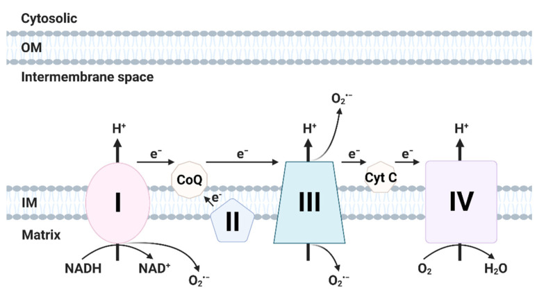
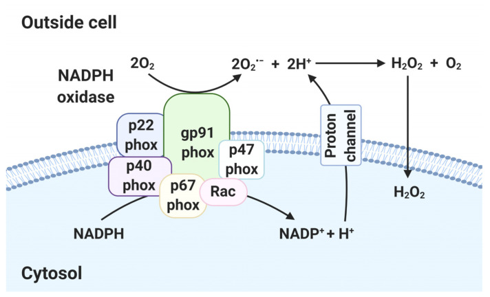
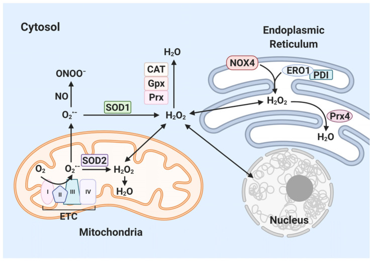
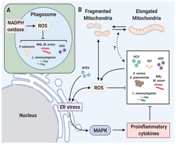
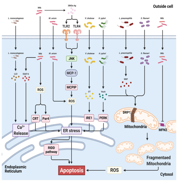
Oxidative stress, particularly reactive oxygen species (ROS), are important for innate immunity against pathogens. ROS directly attack pathogens, regulate and amplify immune signals, induce autophagy and activate inflammation. In addition, production of ROS by pathogens affects the endoplasmic reticulum (ER) and mitochondria, leading to cell death. However, it is unclear how ROS regulate host defense mechanisms. This review outlines the role of ROS during intracellular pathogen infection, mechanisms of ROS production and regulation of host defense mechanisms by ROS. Finally, the interaction between microbial pathogen-induced ROS and the ER and mitochondria is described.

Keywords: ER stress; ROS; bacteria; infection; mitochondria; oxidative stress; pathogen.


Conflict of interest statement

The authors declare no conflict of interest.

Figures

Figure 1
Figure 1
Generation of ROS in electron transport chain (ETC). The ETC is located in the mitochondrial inner membrane (IM). Complex I and II supply electrons to coenzyme Q (CoQ; ubiquinone). Sequentially, electrons are transferred from CoQ to Complex III, cytochrome c (Cyt c) and Complex IV. Oxidative stress is generated during electron transfer. (Created with BioRender.com accessed on 29 March 2021).
Figure 2
Figure 2
Generation of ROS in NADPH oxidase (NOX). In NOX, electrons of cytoplasmic NADPH are transferred to extracellular oxygen via gp91phox, forming O2•−. Other subunits (p22phox, p40phox, p47phox, p67phox and Rac) are responsible for NOX stabilization and regulation. The proton channel transfers protons extracellularly. O2•− is converted to H2O2 and diffuses through lipid membranes into the intracellular space. (Created with BioRender.com accessed on 29 March 2021).
Figure 3
Figure 3
Generation of ROS in cytosol. Superoxide (O2•−, precursor of ROS) is generated in mitochondria and endoplasmic reticulum. O2•− produced in the mitochondrial ETC is dismutated into H2O2 by superoxide dismutase (SOD) 1/2. H2O2 is converted to water by catalase (CAT), glutathione peroxidases (Gpx) and peroxiredoxins (Prx). O2 reacts with NO to produce ONOO. Inside the ER, ER oxidoreductin 1 (ERO1)/protein disulfide isomerase (PDI) and NADPH oxidase 4 (NOX4) produce H2O2. Prx4 reduces H2O2 to water via a catalytic cysteine residue. (Created with BioRender.com accessed on 29 March 2021).
Figure 4
Figure 4
Relationship between ROS and pathogens. Pathogen elimination via ROS-mediated immune signaling. (A) Pathogens are recognized by PRRs, phagocytized and digested. NOX is assembled and activated to produce ROS in the phagosomal membrane, resulting in elimination of the pathogen. (B) ER stress and mitochondrial fragmentation induce ROS production. ROS-induced ER stress boosts proinflammatory cytokine production via MAPK, resulting in microbial killing. Illustration of pathogen-mediated regulation of immune signaling via ROS production. HCV, hepatitis C virus; JEV, Japanese encephalitis virus; HSV, herpes simplex virus 1; BVDV, bovine viral diarrhea virus; S, aureus, Staphylococcus aureus; S. pneumoniae Streptococcus pneumoniae; M. tuberculosis, Mycobacterium tuberculosis; M. avium, Mycobacterium avium; L. monocytogenes, Listeria monocytogenes; F. tularensis, Francisella tularensis. (Created with BioRender.com accessed on 29 March 2021).
Figure 5
Figure 5
Targets of bacteria in ER and mitochondria during infection. Mtb induces ER stress-mediated apoptosis via ROS, calreticulin (CRT), Par-4 (Par4) and Ca2+ release. M. avium increases ROS-mediated ER stress, leading to activation of the RIDD pathway. Cholera toxin (CT) of V. cholera phosphorylates IRE1. Helicobacter pylori secretes VacA, leading to activation of PERK and DRP1. L pneumophila and S. flexneri induce mitochondrial fragmentation in a DRP1-dependent manner. Mtb also triggers mitochondrial fragmentation by inhibiting MFN2. Mtb, Mycobacterium tuberculosis; M. avium, Mycobacterium avium; L. monocytogenes, Listeria monocytogenes; H. pylori, Helicobacter pylori; V. cholera, Vibrio cholera; L pneumophila, Legionella pneumophila; S. flexneri, Shigella flexneri. (Created with BioRender.com accessed on 29 March 2021).

References

    1. Moghadam Z.M., Henneke P., Kolter J. From flies to men: ROS and the NADPH oxidase in phagocytes. Front. Cell Dev. Biol. 2021;9:628991. doi: 10.3389/fcell.2021.628991. - DOI - PMC - PubMed
    1. Herb M., Schramm M. Functions of ROS in macrophages and antimicrobial immunity. Antioxidants. 2021;10:313. doi: 10.3390/antiox10020313. - DOI - PMC - PubMed
    1. Coleman J.W. Nitric oxide in immunity and inflammation. Int. Immunopharmacol. 2001;1:1397–1406. doi: 10.1016/S1567-5769(01)00086-8. - DOI - PubMed
    1. Mayer B., Hemmens B. Biosynthesis and action of nitric oxide in mammalian cells. Trends Biochem. Sci. 1997;22:477–481. doi: 10.1016/S0968-0004(97)01147-X. - DOI - PubMed
    1. Nathan C., Xie Q.W. Nitric oxide synthases: Roles, tolls, and controls. Cell. 1994;78:915–918. doi: 10.1016/0092-8674(94)90266-6. - DOI - PubMed

LinkOut - more resources