

In Vitro Evaluation of Antiproliferative Properties of Novel Organotin(IV) Carboxylate Compounds with Propanoic Acid Derivatives on a Panel of Human Cancer Cell Lines
- PMID: 34071809
- PMCID: PMC8198038
- DOI: 10.3390/molecules26113199
In Vitro Evaluation of Antiproliferative Properties of Novel Organotin(IV) Carboxylate Compounds with Propanoic Acid Derivatives on a Panel of Human Cancer Cell Lines
Abstract
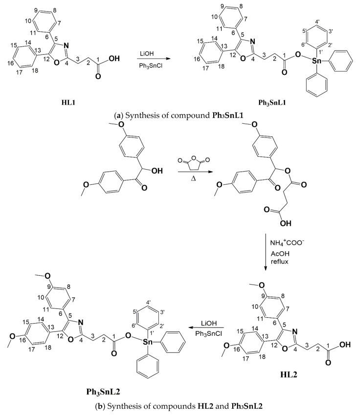
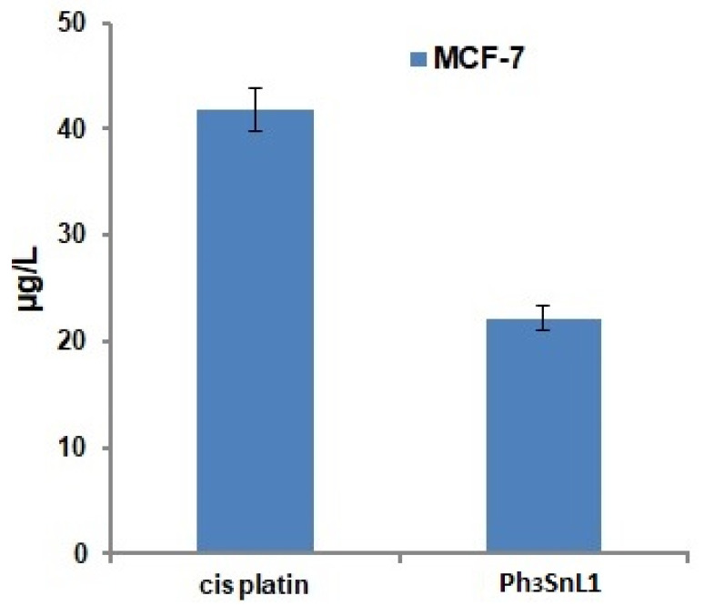
The synthesis of novel triphenyltin(IV) compounds, Ph3SnLn (n = 1-3), with oxaprozin (3-(4,5-diphenyloxazol-2-yl)propanoic acid), HL1, and the new propanoic acid derivatives 3-(4,5-bis(4-methoxylphenyl)oxazol-2-yl)propanoic acid, HL2, and 3-(2,5-dioxo-4,4-diphenylimidazolidin-1-yl)propanoic acid, HL3, has been performed. The ligands represent commercial drugs or their derivatives and the tin complexes have been characterized by standard analytical methods. The in vitro antiproliferative activity of both ligands and organotin(IV) compounds has been evaluated on the following tumour cell lines: human prostate cancer (PC-3), human colorectal adenocarcinoma (HT-29), breast cancer (MCF-7), and hepatocellular cancer (HepG2), as well as on normal mouse embryonic fibroblast cells (NIH3T3) with the aid of MTT (3-(4,5-dimethylthiazol-2-yl)-2,5-12 diphenyltetrazolium bromide) and CV (crystal violet) assays. Contrary to the inactive ligand precursors, all organotin(IV) carboxylates showed very good activity with IC50 values ranging from 0.100 to 0.758 µM. According to the CV assay (IC50 = 0.218 ± 0.025 µM), complex Ph3SnL1 demonstrated the highest cytotoxicity against the caspase 3 deficient MCF-7 cell line. Inductively coupled plasma mass spectrometry (ICP-MS) analysis indicated a two-fold lower concentration of tin in MCF-7 cells in comparison to platinum. To investigate the mechanism of action of the compound Ph3SnL1 on MCF-7 cells, morphological, autophagy and cell cycle analysis, as well as the activation of caspase and ROS/RNS and NO production, has been performed. Results suggest that Ph3SnL1 induces caspase-independent apoptosis in MCF-7 cells.
Keywords: ICP-MS; apoptosis; breast cancer; cytotoxicity; triphenyltin(IV).
Conflict of interest statement
The authors declare no conflict of interest. The funders had no role in the design of the study; in the collection, analyses, or interpretation of data; in the writing of the manuscript; or in the decision to publish the results.
Figures


References
-
- Ksouri R. Food components and diet habits: Chief factors of cancer development (Review) Food Qual. Saf. 2019;3:227–231. doi: 10.1093/fqsafe/fyz021. - DOI
-
- Kritchenkov A.S., Stanishevskii Y.M., Skorik Y.A. Search for new drugs design and antitumour activity of platinum complexes. Pharm. Chem. J. 2019;53:6–14. doi: 10.1007/s11094-019-01947-8. - DOI
MeSH terms
Substances
LinkOut - more resources
Full Text Sources
Medical
Research Materials
Miscellaneous
