

Nigella and Milk Thistle Seed Oils: Potential Cytoprotective Effects against 7β-Hydroxycholesterol-Induced Toxicity on SH-SY5Y Cells
- PMID: 34071950
- PMCID: PMC8229989
- DOI: 10.3390/biom11060797
Nigella and Milk Thistle Seed Oils: Potential Cytoprotective Effects against 7β-Hydroxycholesterol-Induced Toxicity on SH-SY5Y Cells
Abstract
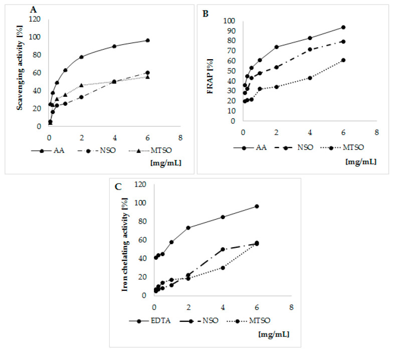
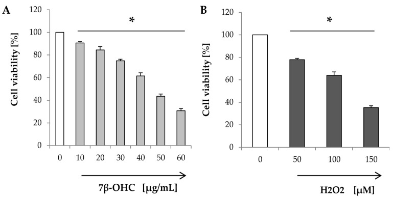
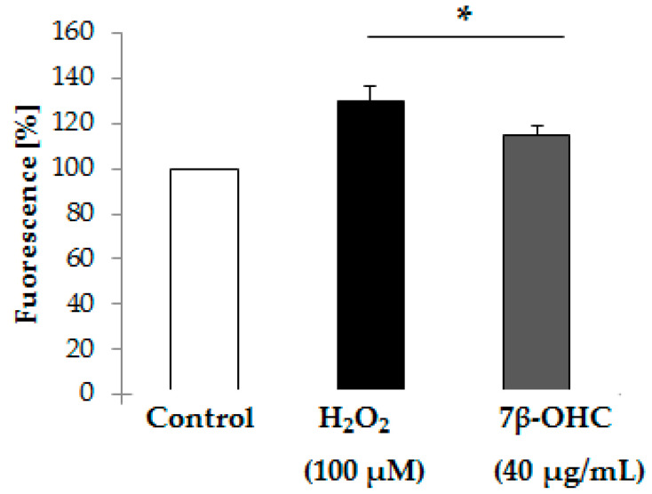
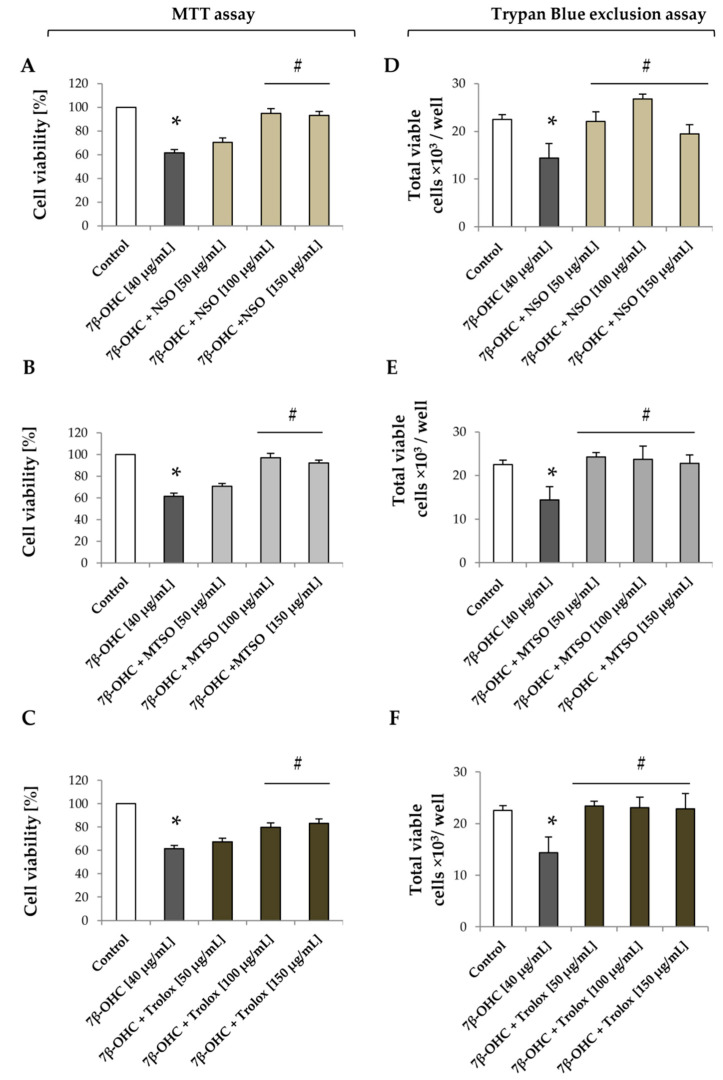
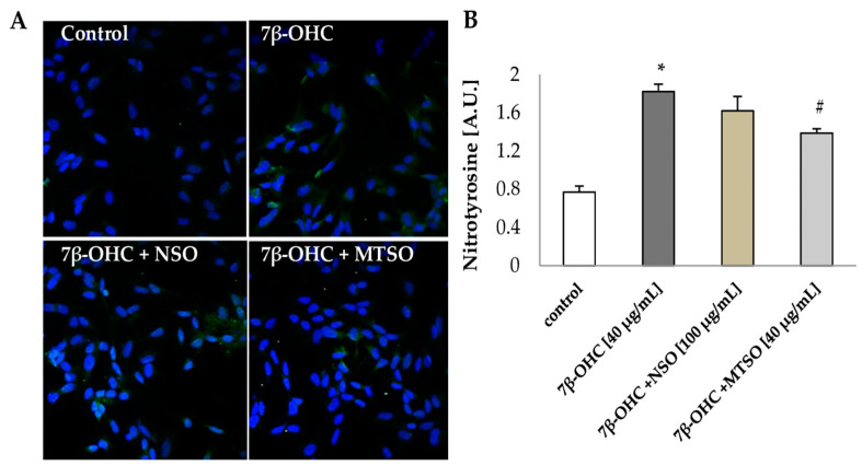
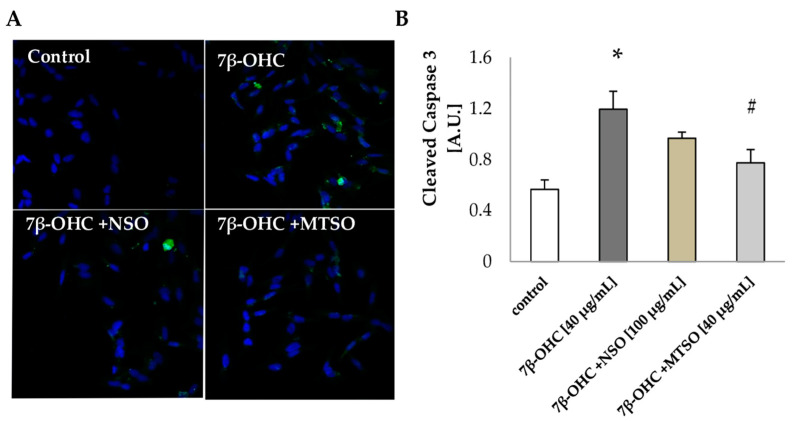
Oxysterols are assumed to be the driving force behind numerous neurodegenerative diseases. In this work, we aimed to study the ability of 7β-hydroxycholesterol (7β-OHC) to trigger oxidative stress and cell death in human neuroblastoma cells (SH-SY5Y) then the capacity of Nigella sativa and Milk thistle seed oils (NSO and MTSO, respectively) to oppose 7β-OHC-induced side effects. The impact of 7β-OHC, associated or not with NSO or MTSO, was studied on different criteria: cell viability; redox status, and apoptosis. Oxidative stress was assessed through the intracellular reactive oxygen species (ROS) production, levels of enzymatic and non-enzymatic antioxidants, lipid, and protein oxidation products. Our results indicate that 7β-OHC (40 µg/mL) exhibit pr-oxidative and pro-apoptotic activities shown by a decrease of the antioxidant enzymatic activities and an increase of ROS production, lipid, and protein oxidation end products as well as nitrotyrosine formation and caspase 3 activation. However, under the pre-treatment with NSO, and especially with MTSO (100 µg/mL), a marked attenuation of oxidative damages was observed. Our study suggests harmful effects of 7β-OHC consisting of pro-oxidative, anti-proliferative, and pro-apoptotic activities that may contribute to neurodegeneration. NSO and especially MTSO showed potential cytoprotection against the cytotoxicity of 7β-OHC.
Keywords: 7β-hydroxycholesterol; antioxidants; antioxidants enzymes; cellular oxidative stress; neuroblastoma cells; neurodegenertion.
Conflict of interest statement
The authors declare no conflict of interest.
Figures
Similar articles
-
Protective effects of milk thistle (Sylibum marianum) seed oil and α-tocopherol against 7β-hydroxycholesterol-induced peroxisomal alterations in murine C2C12 myoblasts: Nutritional insights associated with the concept of pexotherapy.Steroids. 2022 Jul;183:109032. doi: 10.1016/j.steroids.2022.109032. Epub 2022 Apr 4. Steroids. 2022. PMID: 35381271
-
Biotin attenuation of oxidative stress, mitochondrial dysfunction, lipid metabolism alteration and 7β-hydroxycholesterol-induced cell death in 158N murine oligodendrocytes.Free Radic Res. 2019 May;53(5):535-561. doi: 10.1080/10715762.2019.1612891. Free Radic Res. 2019. PMID: 31039616
-
7β-hydroxycholesterol-induced cell death, oxidative stress, and fatty acid metabolism dysfunctions attenuated with sea urchin egg oil.Biochimie. 2018 Oct;153:210-219. doi: 10.1016/j.biochi.2018.06.027. Epub 2018 Jul 9. Biochimie. 2018. PMID: 30003930
-
Attenuation of 7-ketocholesterol- and 7β-hydroxycholesterol-induced oxiapoptophagy by nutrients, synthetic molecules and oils: Potential for the prevention of age-related diseases.Ageing Res Rev. 2021 Jul;68:101324. doi: 10.1016/j.arr.2021.101324. Epub 2021 Mar 24. Ageing Res Rev. 2021. PMID: 33774195 Review.
-
Cytoprotective activities of representative nutrients from the Mediterranean diet and of Mediterranean oils against 7-ketocholesterol- and 7β-hydroxycholesterol-induced cytotoxicity: Application to age-related diseases and civilization diseases.Steroids. 2022 Nov;187:109093. doi: 10.1016/j.steroids.2022.109093. Epub 2022 Aug 24. Steroids. 2022. PMID: 36029811 Review.
Cited by
-
Characterization of Silybum marianum and Silybum eburneum seed oils: Phytochemical profiles and antioxidant properties supporting important nutritional interests.PLoS One. 2024 Jun 14;19(6):e0304021. doi: 10.1371/journal.pone.0304021. eCollection 2024. PLoS One. 2024. PMID: 38875282 Free PMC article.
-
The Dual Role of Amyloid Beta-Peptide in Oxidative Stress and Inflammation: Unveiling Their Connections in Alzheimer's Disease Etiopathology.Antioxidants (Basel). 2024 Oct 8;13(10):1208. doi: 10.3390/antiox13101208. Antioxidants (Basel). 2024. PMID: 39456461 Free PMC article. Review.
References
-
- Testa G., Staurenghi E., Zerbinati C., Gargiulo S., Iuliano L., Giaccone G., Fantò F., Poli G., Leonarduzzi G., Gamba P. Changes in Brain Oxysterols at Different Stages of Alzheimer’s Disease: Their Involvement in Neuroinflammation. Redox Biol. 2016;10:24–33. doi: 10.1016/j.redox.2016.09.001. - DOI - PMC - PubMed
-
- Hammouda S., Ghzaiel I., Khamlaoui W., Hammami S., Mhenni S.Y., Samet S., Hammami M., Zarrouk A. Genetic variants in FADS1 and ELOVL2 increase level of arachidonic acid and the risk of Alzheimer’s disease in the Tunisian population. Prostaglandins Leukot. Essent. Fatty Acids. 2020;160:102159. doi: 10.1016/j.plefa.2020.102159. - DOI - PubMed
Publication types
MeSH terms
Substances
LinkOut - more resources
Full Text Sources
Research Materials
