. 2021 May 27;8(6):92.
doi: 10.3390/vetsci8060092.

Biplanar High-Speed Fluoroscopy of Pony Superficial Digital Flexor Tendon (SDFT)-An In Vivo Pilot Study

Affiliations

Biplanar High-Speed Fluoroscopy of Pony Superficial Digital Flexor Tendon (SDFT)-An In Vivo Pilot Study

Franziska C Wagner et al. Vet Sci. .

Abstract

The superficial digital flexor tendon (SDFT) is the most frequently injured structure of the musculoskeletal system in sport horses and a common cause for early retirement. This project's aim was to visualize and measure the strain of the sound, injured, and healing SDFTs in a pony during walk and trot. For this purpose, biplanar high-speed fluoroscopic kinematography (FluoKin), as a high precision X-ray movement analysis tool, was used for the first time in vivo with equine tendons. The strain in the metacarpal region of the sound SDFT was 2.86% during walk and 6.78% during trot. When injured, the strain increased to 3.38% during walk and decreased to 5.96% during trot. The baseline strain in the mid-metacarpal region was 3.13% during walk and 6.06% during trot and, when injured, decreased to 2.98% and increased to 7.61%, respectively. Following tendon injury, the mid-metacarpal region contributed less to the overall strain during walk but showed increased contribution during trot. Using this marker-based FluoKin technique, direct, high-precision, and long-term strain measurements in the same individual are possible. We conclude that FluoKin is a powerful tool for gaining deeper insight into equine tendon biomechanics.

Keywords: XROMM; collagenase; equine; gait; horse; strain; tendinopathy.


Conflict of interest statement

The authors declare no conflict of interest.

Figures
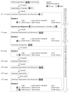
Figure 1
Figure 1
Time schedule of the experimental set-up. Additionally, short clinical examinations were carried out every day within the clinic’s routine.
Figure 2
Figure 2
Computed tomography (CT), FluoKin, and ultrasonographic images of the left forelimb of a pony with implanted tantalum beads. (a) Two tendon markers with metal streak artifact in the CT image, left is dorsal; (b) FluoKin image of the distal forelimb with four tendon markers in the SDFT and five bone markers in the third metacarpal bone; (c) longitudinal and transversal ultrasound image with acoustic shadowing and reverberation artifact due to the implanted beads, left is distal. Arrows mark the implanted beads.
Figure 3
Figure 3
Implantation of the tantalum beads. (a) Left forelimb, lateral (left) and medial (right) view; yellow: bone markers, red: tendon markers with intermarker distances M1 to M4 (#) and M2 to M3 (*); blue: collagenase. (b) Implantation instrument for tendon markers (separated and mounted).
Figure 4
Figure 4
Ultrasonographic image of the left forelimb of a pony, left is lateral: (a) acute (*) and (b) healing (#) tendon lesion of the SDFT (9 weeks after injection of collagenase).
Figure 5
Figure 5
Bar graph of the measured strain of the SDFT of both forelimbs during walk and trot between the implanted tendon markers. Blue arrows: strain behavior in the sound SDFT (right forelimb and “native”). Red dotted arrows: alternated strain behavior compared to “native” and measurements within the right (sound) forelimb. # indicates nonsignificant differences between corresponding measurements in the right, not-insulted forelimb.
Figure 6
Figure 6
Bar graph of the length variation of the tendon markers of both forelimbs during walk and trot. # indicates nonsignificant differences between corresponding amplitudes in the right, not-insulted forelimb.

Similar articles

Cited by

References

    1. Williams R.B., Harkins L.S., Hammond C.J., Wood J.L. Racehorse injuries, clinical problems and fatalities recorded on British racecourses from flat racing and National Hunt racing during 1996, 1997 and 1998. Equine Vet. J. 2001;33:478–486. doi: 10.2746/042516401776254808. - DOI - PubMed
    1. Smith R.K.W., Birch H.L., Goodman S., Heinegård D., Goodship A.E. The influence of ageing and exercise on tendon growth and degeneration–Hypotheses for the initiation and prevention of strain-induced tendinopathies. Comp. Biochem. Physiol. Part. A Mol. Integr. Physiol. 2002;133:1039–1050. doi: 10.1016/S1095-6433(02)00148-4. - DOI - PubMed
    1. Butcher M.T., Hermanson J.W., Ducharme N.G., Mitchell L.M., Soderholm L.V., Bertram J.E.A. Superficial digital flexor tendon lesions in racehorses as a sequela to muscle fatigue: A preliminary study. Equine Vet. J. 2007;39:540–545. doi: 10.2746/042516407X212475. - DOI - PubMed
    1. Patterson-Kane J.C., Firth E.C. The pathobiology of exercise-induced superficial digital flexor tendon injury in Thoroughbred racehorses. Vet. J. 2009;181:79–89. doi: 10.1016/j.tvjl.2008.02.009. - DOI - PubMed
    1. Wollenman P., McMahon P.J., Knapp S., Ross M.W. Lameness in the Polo Pony. In: Ross M.W., Dyson S.J., editors. Diagnosis and Management of Lameness in the Horse. 2nd ed. Elsevier/Saunders; St. Louis, MO, USA: 2011. pp. 1149–1164.

LinkOut - more resources