. 2021 May 27;10(6):861.
doi: 10.3390/antiox10060861.

Genome-Wide Screening of Oxidizing Agent Resistance Genes in Escherichia coli

Affiliations

Genome-Wide Screening of Oxidizing Agent Resistance Genes in Escherichia coli

Hao Chen et al. Antioxidants (Basel). .

Abstract

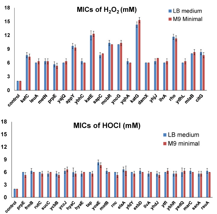
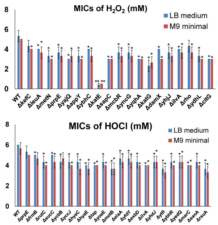
The use of oxidizing agents is one of the most favorable approaches to kill bacteria in daily life. However, bacteria have been evolving to survive in the presence of different oxidizing agents. In this study, we aimed to obtain a comprehensive list of genes whose expression can make Escherichiacoli cells resistant to different oxidizing agents. For this purpose, we utilized the ASKA library and performed a genome-wide screening of ~4200 E. coli genes. Hydrogen peroxide (H2O2) and hypochlorite (HOCl) were tested as representative oxidizing agents in this study. To further validate our screening results, we used different E. coli strains as host cells to express or inactivate selected resistance genes individually. More than 100 genes obtained in this screening were not known to associate with oxidative stress responses before. Thus, this study is expected to facilitate both basic studies on oxidative stress and the development of antibacterial agents.

Keywords: AKSA library; genome-wide screening; hydrogen peroxide; hypochlorite; oxidative stress.


Conflict of interest statement

The authors declare no conflict of interest.

Figures

Figure 1
Figure 1
Bioinformatical analyses of H2O2-resistance genes. The subcellular localization of proteins was obtained from the EcoCyc E. coli Database. The identified proteins were classified into functional categories according to their annotated functions in the UniProt-GOA Database and analyzed by DAVID Bioinformatics Resources. Protein–protein functional interaction networks were analyzed with the STRING database. A high-resolution interaction map is shown in Figure S1.
Figure 2
Figure 2
Bioinformatical analyses of HOCl-resistance genes. The subcellular localization of proteins was obtained from the EcoCyc E. coli Database. The identified proteins were classified into functional categories according to their annotated functions in the UniProt-GOA Database and analyzed by DAVID Bioinformatics Resources. Protein–protein functional interaction networks were analyzed with the STRING database. A high-resolution interaction map is shown in Figure S2.
Figure 3
Figure 3
MICs of selected genes overexpressed in MG1655. MIC determination was performed by varying the concentration of the oxidizing agents, with 1 mM increments from 1 to 20 mM in the LB medium or the M9 minimal medium. The lowest concentration at which bacteria could not grow was recorded as the corresponding MIC. Each strain was tested in three biological replicates. All the differences between MICs of the candidate genes and those of the control were highly significant (p < 0.001).
Figure 4
Figure 4
MICs of selected genes inactivated in BW25113 cells from the Keio collection. MIC determination was performed by varying the concentration of the oxidizing agents, with 1 mM increments from 1 to 10 mM for both H2O2 and HOCl and 0.25 mM increments from 0 to 1 mM (if MICs were lower than 1 mM) in LB medium or M9 minimal medium. The lowest concentration at which bacteria could not grow was recorded as the corresponding MIC. Each strain was tested in three biological replicates. Significant differences (p < 0.05) are marked with *, and highly significant differences (p < 0.001) are marked with **.

Similar articles

Cited by

References

    1. Ezraty B., Gennaris A., Barras F., Collet J.-F. Oxidative stress, protein damage and repair in bacteria. Nat. Rev. Microbiol. 2017;15:385–396. doi: 10.1038/nrmicro.2017.26. - DOI - PubMed
    1. Imlay J.A. The molecular mechanisms and physiological consequences of oxidative stress: Lessons from a model bacterium. Nat. Rev. Microbiol. 2013;11:443–454. doi: 10.1038/nrmicro3032. - DOI - PMC - PubMed
    1. Dixon S.J., Stockwell B.R. The role of iron and reactive oxygen species in cell death. Nat. Chem. Biol. 2014;10:9–17. doi: 10.1038/nchembio.1416. - DOI - PubMed
    1. Redza-Dutordoir M., Averill-Bates D.A. Activation of apoptosis signalling pathways by reactive oxygen species. Biochim. Et Biophys. Acta Mol. Cell Res. 2016;1863:2977–2992. doi: 10.1016/j.bbamcr.2016.09.012. - DOI - PubMed
    1. Imlay J.A. Where in the world do bacteria experience oxidative stress? Environ. Microbiol. 2019;21:521–530. doi: 10.1111/1462-2920.14445. - DOI - PMC - PubMed

LinkOut - more resources