Review
. 2021 May 27;26(11):3228.
doi: 10.3390/molecules26113228.

The Peptide Functionalized Inorganic Nanoparticles for Cancer-Related Bioanalytical and Biomedical Applications

Affiliations
Review

The Peptide Functionalized Inorganic Nanoparticles for Cancer-Related Bioanalytical and Biomedical Applications

Xiaotong Li et al. Molecules. .

Abstract

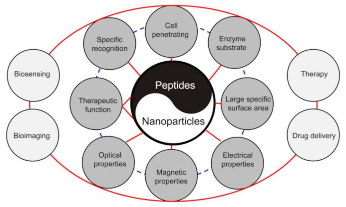
In order to improve their bioapplications, inorganic nanoparticles (NPs) are usually functionalized with specific biomolecules. Peptides with short amino acid sequences have attracted great attention in the NP functionalization since they are easy to be synthesized on a large scale by the automatic synthesizer and can integrate various functionalities including specific biorecognition and therapeutic function into one sequence. Conjugation of peptides with NPs can generate novel theranostic/drug delivery nanosystems with active tumor targeting ability and efficient nanosensing platforms for sensitive detection of various analytes, such as heavy metallic ions and biomarkers. Massive studies demonstrate that applications of the peptide-NP bioconjugates can help to achieve the precise diagnosis and therapy of diseases. In particular, the peptide-NP bioconjugates show tremendous potential for development of effective anti-tumor nanomedicines. This review provides an overview of the effects of properties of peptide functionalized NPs on precise diagnostics and therapy of cancers through summarizing the recent publications on the applications of peptide-NP bioconjugates for biomarkers (antigens and enzymes) and carcinogens (e.g., heavy metallic ions) detection, drug delivery, and imaging-guided therapy. The current challenges and future prospects of the subject are also discussed.

Keywords: biosensing nanoplatform; cancer; inorganic nanoparticle; nanomedicine; peptide ligand.


Conflict of interest statement

The authors declare no conflict of interest.

Figures

Figure 1
Figure 1
Schematic illustration of the effects of properties of peptide functionalized NPs on bioanalytical and biomedical areas including biosensing, bioimaging, drug delivery, and therapy. Combination of the biological properties of peptides and unique physicochemical properties of NPs can generate excellent nanosensing platforms with high sensitivity and specificity and nanotheranostics with high tumor-targeting capacity.
Figure 2
Figure 2
Schematic representation of amide bond formation between the KPQPRPLS peptide and OEG NPs by using EDC and Sulfo-NHS. The degree of peptide coupling as well as the colloidal stability of PEP-OEP NPs are strongly dependent on the experimental conditions (such as EDC and Sulfo-NHS concentrations, peptide concentration, reaction time, and reaction buffer) (Adapted from Bartczak and Kanaras 2011 [58], Copyright 2011 The American Chemical Society and reproduced with permission.).
Figure 3
Figure 3
AuNP-(EK)3 nanoprobe-based colorimetric detection of Ni2+. (a) Schematic illustration of AuNP-(EK)3 nanoprobe preparation and detection principle of Ni2+, (b) UV–visible absorption spectra of AuNP-(EK)3 at different concentrations of Ni2+ (0–50 μM), (c) linear calibration plot of AuNP-(EK)3 versus Ni2+ concentrations in the range of 60–160 nM, (d) selectivity of AuNP-(EK)3 toward different metal ion species. The zwitterionic region of the (EK)3-peptide can bind to Ni2+ due to the presence of an -NH2 or -COOH group and the unfilled d-orbital of Ni2+, leading to the aggregation of AuNPs. As a result, in the presence of Ni2+, the color of the AuNP-(EK)3 solution is changed from red to purple. (Adapted from Parnsubsakul et al. 2018 [79], Copyright 2018 The Royal Society of Chemistry and reproduced with permission.).
Figure 4
Figure 4
(a) Comparison of the size of the membrane with respect to a 50 cents coin, and (b) photograph and (c) schematics of the assay with peptide functionalized AuNPs on PVDF membrane, which yields a change in color from reddish to violet, due to aggregation induced by MMP-7. The MMP-7 peptide substrate functionalized AuNPs were deposited on PVDF membrane. The AuNPs aggregate on PVDF membrane because of increased electrostatic interparticle attraction upon proteolysis by MMP-7. (Adapted from Goyal et al. 2020 [98], Copyright 2019 Elsevier B.V. and reproduced with permission.).
Figure 5
Figure 5
The schematic representation of UCNP-based FRET sensing platform for the detection of caspase-9 activity both in vitro and in vivo by using UCNPs as the energy donor and Cy5 as the energy acceptor. The Cy5 labelled peptide with specific motif LEHD for caspase-9 cleavage were conjugated with carboxyl modified UCNP@SiO2 through covalent attachment. (Adapted from Liu et al. 2019 [114], Copyright 2019 Elsevier B.V. and reproduced with permission.).
Figure 6
Figure 6
Schematic illustration for the synthesis of RGD and Cys functionalized Gd(OF)3: Ce, Tb nanocrystals and their targeted imaging. The oleylamine capped nanocrystals were modified by polyacrylic acid (PAA) to form the PAA capped Gd(OF)3: 45%Ce, 15%Tb nanocrystals (PAA@ Gd(OF)3: 45%Ce, 15%Tb) through a ligand exchange method. RGD peptide and cysteine were conjugated to the nanocrystals surface by EDC/NHS chemistry (Adapted from Yan et al. 2015 [154], Copyright 2015 Elsevier Ltd. and reproduced with permission.).
Figure 7
Figure 7
Antiangiogenic cancer therapy by AF@AuNCs. (a) Schematic illumination of Anti-Flt1 peptide templated AF@AuNCs for antitumor angiogenesis, (b) inhibition of CAM angiogenesis after incubation with AF, AF@AuNCs, and GSH@AuNCs at a concentration of 100 μg/mL and the incubation time was 48 h, and (c) comparison of the number of vessels in the AF, AF@AuNCs, and GSH@AuNCs after incubation for 48 h at a concentration of 100 μg/mL in the CAM model. AF@AuNCs were prepared by mixing AF with HAuCl4. The AF@AuNCs show enhanced ability in inhibiting angiogenesis in fertilized eggs than those of pure AF and GSH@AuNCs. Data represent means ± SD (n = 3). * p < 0.05 and *** p < 0.001, data with significant difference (Adapted from Li et al. 2021 [181], Copyright 2021 American Chemical Society and reproduced with permission.).
Figure 8
Figure 8
Schematic representation of MET-induced stromal depletion for enhancing the penetration and cathepsin B-triggered release of GEM carried by Fe3O4 NPs in the lysosome of pancreatic ductal adenocarcinoma (PADC) cells. The pHLIP facilitates the internalization of the underlying Fe3O4 NPs by inserting into cell membranes because it can format stable transmembrane α-helix in acidic tumor microenvironment. Once internalized into the cancer cells, GEM will be released in lysosome upon cleavage of its linker (i.e., GFLG peptide) by cathepsin B. In the animal experiments, MET was intraperitoneally injected to deplete the dense stromal barrier of PDAC prior to the injection of the above nanoagents to facilitate the effective delivery of GEM. (Adapted from Han et al. 2020 [205], Copyright 2020 American Chemical Society and reproduced with permission.).
Figure 9
Figure 9
Schematic representation of the cRGD–PLGA–SPIO@DOX multifunctional NPs for targeted tumor therapy and MR imaging. The SPIO and DOX were encapsulated by cRGD peptide-functionalized poly(lactic-coglycolic acid) (cRGD–PLGA) block copolymer. The as-synthesized nanosystem (cRGD–PLGA–SPIO@DOX) exhibited excellent pH-responsive drug release properties under physiological conditions and integrin-targeting ability, which can act as a theranostic agent for MRI-guided cancer therapy (Adapted from Xiao et al. 2019 [229], Copyright 2019 The Royal Society of Chemistry and reproduced with permission.).
Figure 10
Figure 10
(a) Schematic representation of the formation of RGDfC-SeNPs/siRNA, and (b) the main signaling pathway of apoptosis induced by RGDfC-SeNPs/siRNA. The SeNPs were synthesized through reduction of Na2SeO3 by ascorbic acid (Vc), which were functionalized by the cancer-targeting ligand, RGDfC peptide. The siRNA was then loaded on the surface of RGDfC-SeNPs to prepare RGDfC-SeNPs/siRNA. The RGDfC-SeNPs/siRNA can induce cell apoptosis by regulating the Wnt/β-catenin signaling and activation of its downstream target gene (Adapted from Xia et al. 2017 [239], Copyright 2017 The Royal Society of Chemistry and reproduced with permission.).
Figure 11
Figure 11
(a) Schematic depiction for the synthesis of AuNP-DPA and their enrichment in the tumor site. The AuNPs were modified by the C terminus of DPA (DPA-Cys, encapsulated by PLL, and functionalized by RGD-derived peptide RGDDP. (b) The AuNP-DPA can efficiently re-lease DPA to the cytosol through breaking the gold-thiolate bonds by the intracellular reductant, such as GSH. (Adapted from Bian et al. 2018 [257], Copyright 2018 Ivyspring International Pub-lisher and reproduced with permission.).

Similar articles

Cited by

References

    1. Wild C., Weiderpass E., Stewart B. In: World Cancer Report: Cancer Research for Cancer Prevention. Wild C., Weiderpass E., Stewart B., editors. International Agency for Research on Cancer; Lyon, France: 2020.
    1. Rosi N.L., Mirkin C.A. Nanostructures in biodiagnostics. Chem. Rev. 2005;105:1547–1562. doi: 10.1021/cr030067f. - DOI - PubMed
    1. Wang Z.X., Ma L.N. Gold nanoparticle probes. Coordin. Chem. Rev. 2009;253:1607–1618. doi: 10.1016/j.ccr.2009.01.005. - DOI
    1. Zhou W., Gao X., Liu D.B., Chen X.Y. Gold Nanoparticles for In Vitro Diagnostics. Chem. Rev. 2015;115:10575–10636. doi: 10.1021/acs.chemrev.5b00100. - DOI - PMC - PubMed
    1. Trouiller A.J., Hebie S., El Bahhaj F., Napporn T.W., Bertrand P. Chemistry for oncotheranostic gold nanoparticles. Eur. J. Med. Chem. 2015;99:92–112. doi: 10.1016/j.ejmech.2015.05.024. - DOI - PubMed

MeSH terms