. 2021 May 27;22(11):5733.
doi: 10.3390/ijms22115733.

PSA Depletion Induces the Differentiation of Immature Neurons in the Piriform Cortex of Adult Mice

Affiliations

PSA Depletion Induces the Differentiation of Immature Neurons in the Piriform Cortex of Adult Mice

Simona Coviello et al. Int J Mol Sci. .

Abstract

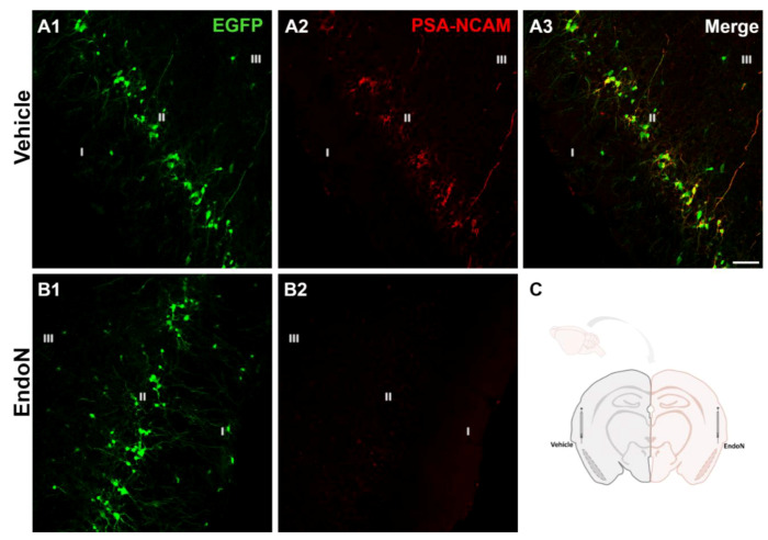
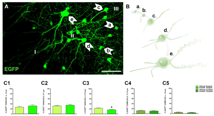
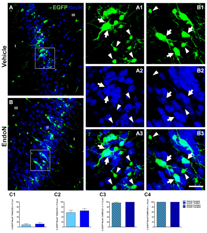
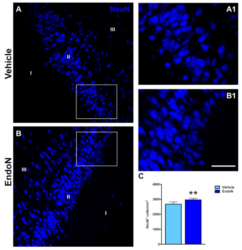
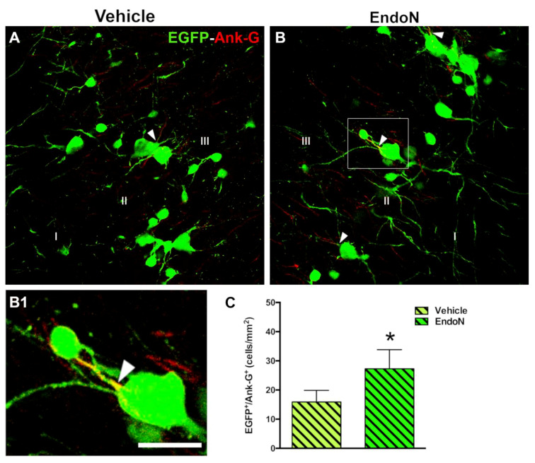
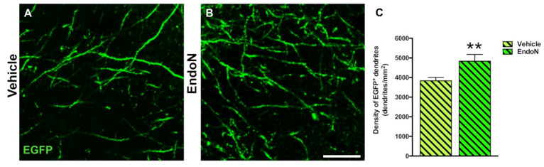
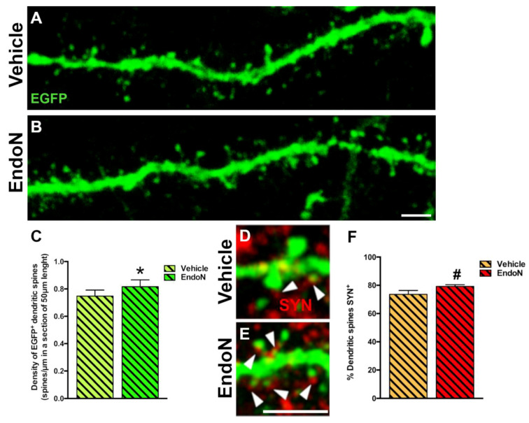
Immature neurons are maintained in cortical regions of the adult mammalian brain. In rodents, many of these immature neurons can be identified in the piriform cortex based on their high expression of early neuronal markers, such as doublecortin (DCX) and the polysialylated form of the neural cell adhesion molecule (PSA-NCAM). This molecule plays critical roles in different neurodevelopmental events. Taking advantage of a DCX-CreERT2/Flox-EGFP reporter mice, we investigated the impact of targeted PSA enzymatic depletion in the piriform cortex on the fate of immature neurons. We report here that the removal of PSA accelerated the final development of immature neurons. This was revealed by a higher frequency of NeuN expression, an increase in the number of cells carrying an axon initial segment (AIS), and an increase in the number of dendrites and dendritic spines on the immature neurons. Taken together, our results demonstrated the crucial role of the PSA moiety in the protracted development of immature neurons residing outside of the neurogenic niches. More studies will be required to understand the intrinsic and extrinsic factors affecting PSA-NCAM expression to understand how the brain regulates the incorporation of these immature neurons to the established neuronal circuits of the adult brain.

Keywords: PSA-NCAM; doublecortin; neuronal maturation; neuronal precursors; olfactory cortex.


Conflict of interest statement

The authors declare no conflict of interest.

Figures

Figure 1
Figure 1
Removal of PSA in the PCX by Endo-N. (A,B) Confocal microscopic images of EGFP (green) and PSA-NCAM (red) immunoreactive neurons in the vehicle (A13) and Endo-N (B1,2) injected hemispheres. Note the complete absence of PSA-NCAM expression in the hemisphere injected with Endo-N. (C) Drawing showing the hemispheres injected with vehicle (left) or Endo-N (right). Scale bar: 70 µm.
Figure 2
Figure 2
Classification of EGFP+ neurons and impact of PSA depletion. (A) Single confocal section showing the distribution of EGFP+ cells in the PCX layer II. Note the different subtypes of cells indicated by arrows. Scale bar = 50 µm (B) Schematic representation showing the different maturational stages of EGFP+ cells: a: ≤ 5 µm; b: 6–7 µm; c: 8–10 µm; d: 11–13 µm; e: ≥ 14 µm (C15) Graphs representing the percentage of the different types and maturational stages of EGFP+ neurons in the PCX layer II. Asterisks indicate statistically significant differences between hemispheres (Vehicle vs. Endo-N) after paired Student’s t-test. (* p ≤ 0.05) Scale bar: 100 µm.
Figure 3
Figure 3
Effects of Endo-N injection on the percentage of EGFP+ cells displaying NeuN+ nuclei. (A,B) Confocal images showing EGFP+ cells (green) and NeuN-expressing cells (blue). (A13,B13) Higher magnifications of the squared areas in A and B. In these insets, tangled and complex EGFP+ cells are indicated by arrowheads and arrows, respectively. Note the presence of a larger number of EGFP+ complex cells labeled with NeuN in the Endo-N hemisphere. (C14) Graphs representing the percentage of EGFP+/NeuN+ neurons classified as tangled or complex cells according to their diameter. Scale bar: 70 µm.
Figure 4
Figure 4
Effects of Endo-N injection on the density of NeuN-expressing cells. (A,B) Single confocal sections showing the expression of NeuN in the PCX. (A1,B1) are higher magnifications of the squared areas in (A,B), which show the different densities of NeuN immunoreactive nuclei in the PCX layer II. (C) Graph showing the effect of Endo-N injection on the density of NeuN immunoreactive nuclei in the PCX layer II. Asterisks indicate statistically significant differences between hemispheres (vehicle vs. Endo-N) after paired Student’s t-test. (** p < 0.01). Scale bar represents 70 µm for (A,B) and 120 µm for (A1,B1).
Figure 5
Figure 5
Effects of Endo-N injection on the density of EGFP+ complex cells displaying an AIS. (A,B) Panoramic views of the PCX layer II with EGFP+ cells (green) showing axon initial segments (AIS) (red) identified by the expression of the protein Ank-G. Note the presence of a larger number of EGFP+ complex cells displaying an AIS in the Endo-N hemisphere. (B1) Higher magnification of the squared area in B. An arrowhead indicates the AIS. (C) Graph showing a higher density of EGFP+ complex cells expressing Ank-G in the Endo-N hemisphere. Asterisks indicate statistically significant differences between hemispheres (Vehicle vs. Endo-N) after paired Student’s t-test. (* p ≤ 0.05) Scale bar represents 30 µm for A and B, and 55 µm for B1.
Figure 6
Figure 6
Confocal analysis of the density of EGFP+ dendrites in the PCX layer I. (A,B) Single confocal planes showing dendritic processes in the vehicle (A) and Endo-N (B) hemispheres. (C) Graph showing a higher density of dendrites in the Endo-N versus the vehicle hemisphere. Asterisks indicate statistically significant differences between hemispheres (Vehicle vs. Endo-N) after paired Student’s t-test. (** p < 0.01). Scale bar: 10 µm.
Figure 7
Figure 7
Confocal analysis of the density of dendritic spines in the PCX layer I. (A,B) 2D projections of confocal stacks (20 confocal planes separated by 0.38 µm) of spiny dendrites from the vehicle (A) and Endo-N (B) hemispheres. (C) Graph showing increased the density of dendritic spines in a segment of 50 µm in the Endo-N hemisphere. (D,E) Higher magnification views of the dendritic spines of complex EGFP+ cells in close apposition to puncta expressing the presynaptic markers SYN (red) on their surface. Note the presence of a larger number of puncta expressing SYN (arrowheads) in the Endo-N hemisphere (E) when compared to the vehicle hemisphere (D). (F) Graphs showing the percentage of puncta expressing SYN apposed to EGFP+ spines. Asterisks indicate statistically significant differences between hemispheres (Vehicle vs. Endo-N) after paired Student’s t-test. (* p ≤ 0.05; 0.05 < # p < 0.1). Scale bar: 5 µm (B) and 15 µm (E).
Figure 8
Figure 8
Excitatory and inhibitory puncta in the perisomatic region of EGFP+ complex cells in the PCX layer II. (A,B) 2D projections of single confocal stacks showing VGLUT1+ (A, red), VGAT+ (B, blue) and PV+ (B, red) puncta in the perisomatic region of EGFP+ complex cells (green) in the vehicle (A1,B1) and Endo-N (A2,B2) injected hemispheres. Scale bar: 10 µm.

Similar articles

Cited by

References

    1. Lois C., Alvarez-Buylla A. Long-Distance Neuronal Migration in the Adult Mammalian Brain. Science. 1994;264:1145–1148. doi: 10.1126/science.8178174. - DOI - PubMed
    1. Gage F.H. Mammalian Neural Stem Cells. Science. 2000;287:1433–1438. doi: 10.1126/science.287.5457.1433. - DOI - PubMed
    1. Feliciano D.M., Bordey A., Bonfanti L. Noncanonical Sites of Adult Neurogenesis in the Mammalian Brain. Cold Spring Harb. Perspect Biol. 2015;7 doi: 10.1101/cshperspect.a018846. - DOI - PMC - PubMed
    1. Castillo-Gómez E., Gómez-Climent M.Á., Varea E., Guirado R., Blasco-Ibáñez J.M., Crespo C., Martínez-Guijarro F.J., Nácher J. Dopamine Acting through D2 Receptors Modulates the Expression of PSA-NCAM, a Molecule Related to Neuronal Structural Plasticity, in the Medial Prefrontal Cortex of Adult Rats. Exp. Neurol. 2008;214:97–111. doi: 10.1016/j.expneurol.2008.07.018. - DOI - PubMed
    1. Bonfanti L., Nacher J. New Scenarios for Neuronal Structural Plasticity in Non-Neurogenic Brain Parenchyma: The Case of Cortical Layer II Immature Neurons. Prog. Neurobiol. 2012;98:1–15. doi: 10.1016/j.pneurobio.2012.05.002. - DOI - PubMed