Review
. 2021 May 27;9(6):1154.
doi: 10.3390/microorganisms9061154.

Nebulized Colistin in Ventilator-Associated Pneumonia and Tracheobronchitis: Historical Background, Pharmacokinetics and Perspectives

Collaborators, Affiliations
Review

Nebulized Colistin in Ventilator-Associated Pneumonia and Tracheobronchitis: Historical Background, Pharmacokinetics and Perspectives

Yinggang Zhu et al. Microorganisms. .

Abstract

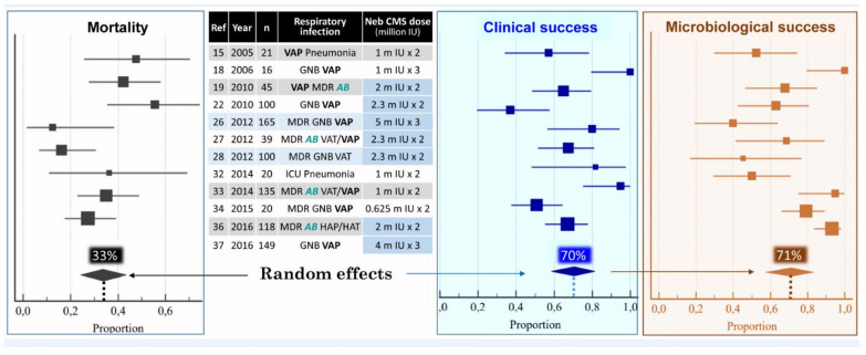
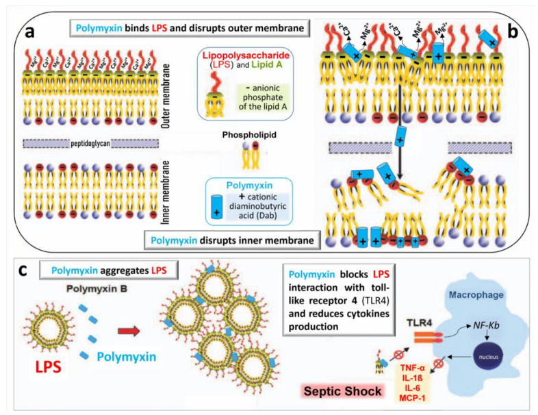
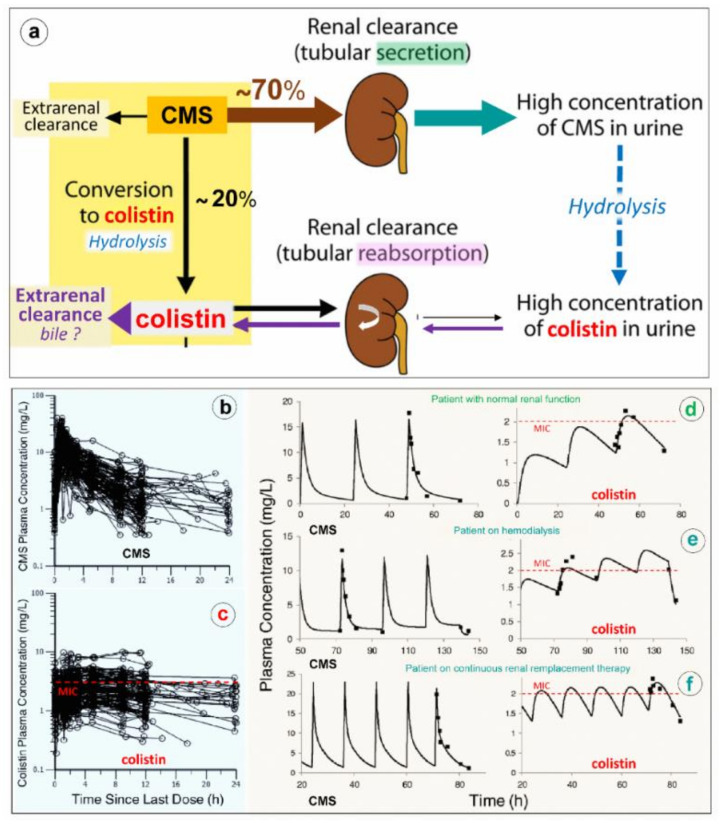
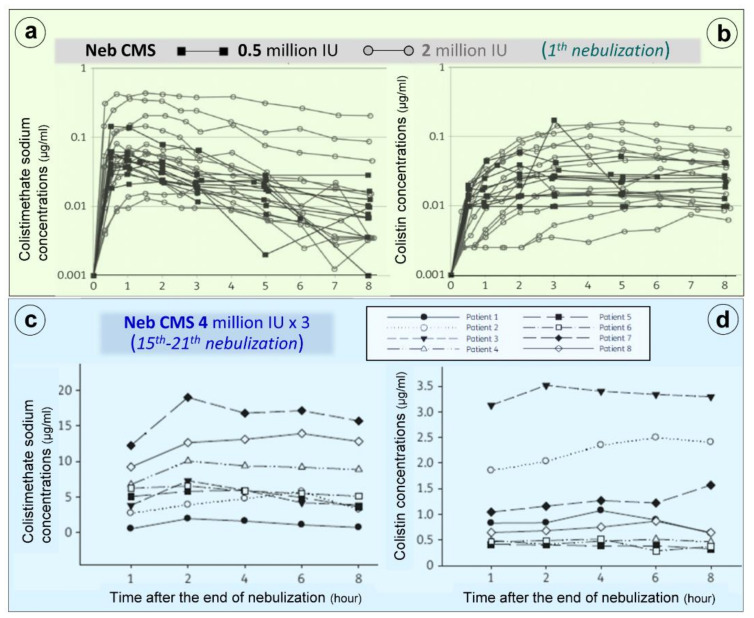
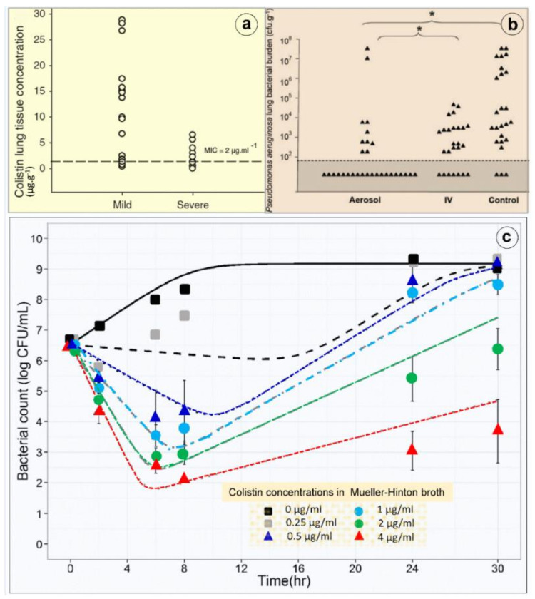
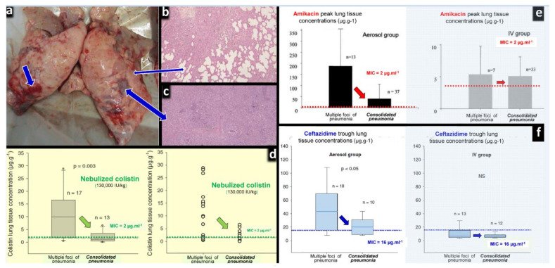
Clinical evidence suggests that nebulized colistimethate sodium (CMS) has benefits for treating lower respiratory tract infections caused by multidrug-resistant Gram-negative bacteria (GNB). Colistin is positively charged, while CMS is negatively charged, and both have a high molecular mass and are hydrophilic. These physico-chemical characteristics impair crossing of the alveolo-capillary membrane but enable the disruption of the bacterial wall of GNB and the aggregation of the circulating lipopolysaccharide. Intravenous CMS is rapidly cleared by glomerular filtration and tubular excretion, and 20-25% is spontaneously hydrolyzed to colistin. Urine colistin is substantially reabsorbed by tubular cells and eliminated by biliary excretion. Colistin is a concentration-dependent antibiotic with post-antibiotic and inoculum effects. As CMS conversion to colistin is slower than its renal clearance, intravenous administration can lead to low plasma and lung colistin concentrations that risk treatment failure. Following nebulization of high doses, colistin (200,000 international units/24h) lung tissue concentrations are > five times minimum inhibitory concentration (MIC) of GNB in regions with multiple foci of bronchopneumonia and in the range of MIC breakpoints in regions with confluent pneumonia. Future research should include: (1) experimental studies using lung microdialysis to assess the PK/PD in the interstitial fluid of the lung following nebulization of high doses of colistin; (2) superiority multicenter randomized controlled trials comparing nebulized and intravenous CMS in patients with pandrug-resistant GNB ventilator-associated pneumonia and ventilator-associated tracheobronchitis; (3) non-inferiority multicenter randomized controlled trials comparing nebulized CMS to intravenous new cephalosporines/ß-lactamase inhibitors in patients with extensive drug-resistant GNB ventilator-associated pneumonia and ventilator-associated tracheobronchitis.

Keywords: colistin; multidrug resistant gram-negative bacteria; nebulized colistimethate sodium; nebulized polymyxin; pharmacodynamics; phramacokinetic; polylyxin resistance; technique of nebulization; ventilator-associated pneumonia; ventilator-associated tracheobronchitis.


Conflict of interest statement

The authors declare receiving consulting fees, unrestricted research grants and equipment research support from Aerogen Ltd., unrestricted research grant, speaker fees, travel reimbursements from Fisher & Paykel, unrestricted research grant form Hamilton medical, consulting fees from La Diffusion Technique Française. M.L. is a consultant for Gilead and Amomed and gave lectures for Aspen and MSD. P.F.L. is a consultant for Adrenomed and Inotrem and received an unrestricted research grant from Aerogen. L.P. declares that Stony Brook University holds patents on targeted antibiotic therapy to intubated patients licensed to InspiRx, Inc. and that she serves as a consultant to InspiRx and is a member of Merck’s Advisory Committee for Gram-negative pneumonias. JRe received grant support from Bayer and served in the advisory board for Bayer and speakers bureau for Norma Helas. A.T. declares participating to the advisory board of Cardeas, Bayer and Polyphor and receiving unrestricted research grants from the three companies. T.W. received grant support from German Research Council, German MInistry of Research and Education, received fees for lectures from AstraZeneca, Basilea, Bayer, GSK, Infectopharm, MSD, Novartis, Pfizer, Roche and declares participating to the advisory board of AstraZeneca, Basilea, Bayer, GSK, Novartis, Pfizer, Roche. The other authors declare no conflict of interest. K.P. received lecture fees from MSD Greece and Pfizer Hellas.

Figures

Figure 1
Figure 1
Pooled analysis of mortality, clinical and microbiological success among 908 patients treated with nebulized colistimethate sodium (CMS) for ventilator-associated pneumonia (VAP) or ventilator-associated tracheobronchitis (VAT) caused by multidrug-resistant (MDR) Gram-negative bacteria (GNB), particularly Acinetobacter baumannii (AB). Squares = proportion in each study; horizontal lines = 95% CI; diamonds = pooled proportion for the 12 studies. Doses of nebulized CMS are expressed in million international unit (IU). Adapted with permission from ref. [44]. Copyright 2017 Elsevier.
Figure 2
Figure 2
Representative computed tomography images obtained in a patient with ventilator-associated pneumonia (VAP) caused by multidrug-resistant Pseudomonas aeruginosa and treated by nebulized colistimethate sodium (CMS) 5 million international units × 3 /24 h for 10 days. A color encoding system identifies normally aerated lung regions (dark grey), poorly aerated lung regions (light grey) and nonaerated consolidated lung regions (red). (a) Contiguous 10 mm thick computed tomography sections obtained before nebulization (day 0) shows bilateral consolidation of lower lobes with disseminated foci of interstitial pneumonia in upper lobes. (b) Ten days later, lung consolidations are partially reaerated, attesting to the clinical efficiency of nebulized CMS monotherapy. (c,d) Computed tomography quantitative assessment of gas volume (aeration) and tissue volume (inflammation/infection) before and after CMS (colistin) nebulization in seven patients with VAP caused by MDR Pseudomonas aeruginosa. Nebulized CMS monotherapy was associated with a significant re-aeration and decrease in inflammation/infection. (e) Changes in Clinical Pulmonary Infection Score in 29 patients with VAP caused by MDR Pseudomonas aeruginosa or Acinetobacter baumannii successfully treated by nebulized CMS (green color) and in 13 patients with VAP caused by MDR Pseudomonas aeruginosa or Acinetobacter baumannii unsuccessfully treated by nebulized CMS (blue color). * indicates p < 0.001 [26].
Figure 3
Figure 3
Chemical structures of polymyxin B (PMB), colistin and colistimethate sodium. (a) PMB is characterized by D-Phenyl and colistin by D-Leuc in position 6. Each antibiotic is a mixture of active components differing by the type of fatty acyl chain linked to the N-terminal diaminobutyric acid (Dab) residue. At physiological pH, Dab are positively charged + and interact with anionic phosphates of lipid A of LPS, thereby disrupting the bacterial outer membrane; colistin A and PMB1 = (S)-6-methyl-octanoic acid; colistin B and PMB2 = (S)-6-methyl-heptanoic acid; colistin C and PMB3 = octanoyl acid; colistin D and PMB4 = heptanoyl acid; colistin E and PMB5 = nonanoyl; colistin F and PMB6 = 3-hydroxy-6- methyloctanoyl acid. Light grey identifies the polar residues of the heptapeptide, light purple the hydrophobic motif within the heptapeptide ring and dark grey the N-terminal fatty acid analogues. (b) Colistimethate sodium, the inactive prodrug of colistin, is prepared from colistin by reaction of the free γ-amino groups of the Dab residues with formaldehyde followed by sodium bisulfite (methanesulfonate moieties). Colistimethate sodium A and B are defined by the fatty acid chain linked to Dab in position 1: (S)-6-methyl octanoic acid for colistimethate A and (S)-6-methyl heptanoic acid for colistimethate B. At physiological pH, methanesulfonate moieties are negatively charged—and Dab cannot interact anymore with anionic phosphates of lipid A of LPS, thereby precluding any bactericidal effect (see Figure 4). Adapted with permission from ref. [54]. Copyright 2009 American Chemical Society.
Figure 4
Figure 4
Schematic representation of the bactericidal action of polymyxins. (a) The outer membrane of the Gram-negative bacterial wall is stabilized by the electrostatic interaction between divalent cations Ca++ and Mg++ and negatively charged phosphodiesters of lipid A of lipopolysaccharide (LPS), thereby creating a permeability barrier against harmful external agents. (b) Polymyxins disrupt the physiological bridges LPS/lipid A. The positively charged Dab interacts electrostatically with negatively charged phospholipids of the LPS, producing the leakage of cellular components through the disrupted bacterial membrane. The mechanisms by which polymyxins disrupt the bacterial inner membrane remain undetermined. (c) Polymyxins aggregate free LPS released from the bacterial wall and block LPS interaction with macrophage receptor, TLR4. The NF-kB pathway is no longer stimulated, and release of pro-inflammatory cytokines such as tumor-necrosing factor-α (TNF-α), interleukins 1β and 6 (IL-1β and IL6) and monocyte chemoattractant protein 1 (MCP-1) is limited, reducing the sepsis severity [26].
Figure 5
Figure 5
Worldwide distribution of mcr-1-producing isolates in humans and animals [63].
Figure 6
Figure 6
Elimination and pharmacokinetics of colistimethate sodium (CMS) and colistin. (a) Schematic representation of elimination pathways for CMS and colistin. Arrow thickness indicates the relative magnitude of each pathway when kidney function is normal. CMS includes all partially methanesulfonated derivatives of colistin. After intravenous administration of CMS, extensive renal excretion occurs, with some of the excreted CMS being converted to colistin within the urinary tract. Colistin is massively reabsorbed by renal tubules and likely excreted in the biliary tract [63]. (b,c) Plasma concentration time profiles of CMS (Figure 6b) and formed colistin (Figure 6c) with 105 critically ill patients who were treated by intravenous CMS for blood stream infection or pneumonia caused by multidrug-resistant Gram-negative bacteria (89 not on renal replacement, 12 on intermittent hemodialysis and 4 on continuous renal replacement therapy). Doses of replacement therapy (df). The dashed line indicates the minimum inhibitory concentrations of susceptible strains [69].
Figure 7
Figure 7
Colistimethate sodium (CMS) and colistin plasma concentrations following nebulization of various doses of CMS. (a,b) Plasma concentrations measured following the initial nebulization of CMS 0.5 and 2 million IU in a series of twelve patients with ventilator-associated pneumonia caused by Gram-negative bacteria [79]. (c,d) Plasma concentrations measured after 2–7 days of CMS nebulization at a dose of 4 million IU three times a day in a series of eight patients with ventilator-associated pneumonia caused by multidrug-resistant Gram-negative bacteria. Plasma concentrations were measured by high-performance liquid chromatography in the 8 h following an individual nebulization [81].
Figure 8
Figure 8
Lung deposition and bactericidal effects of high-dose nebulized colistimethate sodium. (a) Colistin concentrations measured in multiple post-mortem subpleural lung specimens in a series of six anesthetized and mechanically ventilated piglets with inoculation pneumonia caused by Pseudomonas aeruginosa. Colistin concentrations were measured by high-performance liquid chromatography in 17 pulmonary segments with mild pneumonia and moderate loss of lung aeration and in 13 pulmonary segments with severe pneumonia and complete loss of lung aeration (infectious consolidation). The dashed line indicates the minimal inhibitory concentration of the inoculated Pseudomonas aeruginosa [42]. (b) Lung bacterial burden of Pseudomonas aeruginosa measured in post-mortem lung segments in sixteen piglets with massive inoculation pneumonia caused by Pseudomonas aeruginosa. Six received three nebulizations of 100,000 IU/kg colistimethate sodium at 12 h intervals (aerosol), six received four intravenous administrations of 40,000 IU/kg at 8 h intervals (IV) and four did not received any antibiotic (control). Quantitative lung bacteriology was measured in lung segments (triangles) sampled 1 h after the third aerosol in the aerosol group and after the fourth infusion in the intravenous group (IV) and 49 h after the bacterial inoculation in the untreated control group. The grey area indicates the lower limit of quantification for bacterial counts. Asterisk at the top of the figure indicates the statistically significant difference existing between the percentage of lung segments characterized by bacterial counts ranging between 0 and 102 cfu∙g−1 in aerosol and intravenous groups and in aerosol and control groups [42]. (c) Colistin in vitro time–kill curve. An inoculum of 5 × 10⁶ colony forming unit (CFU)/mL of a wild-type Pseudomonas aeruginosa strain was prepared by a suspension of the bacteria from an 18 h logarithmic-growth-phase culture in Mueller–Hinton broth. The experiments were performed in 10 mL glass tubes that were incubated at 37 °C for 18 to 24 h. Colistin was added to obtain concentrations of 0.25, 0.5, 1, 2 and 4 µg/mL (corresponding to 0.5 to eight times the minimal inhibitory concentrations). The bacteria were counted at 0, 2, 6, 8, 24 and 30 h. The limit of quantification was 100 CFU/mL. Four replicates were performed for each concentration. At least one growth control, without added colistin, was included in each experiment. Four replicates were performed for each concentration. Colistin provides a concentration-dependent bacterial killing (means and standard deviations from four replicates and model predicted curves (lines) with mean parameter estimates) [79].
Figure 9
Figure 9
Effects of lung aeration loss on the lung tissue concentrations after the nebulization of high doses of colistin, amikacin and ceftazidime to anesthetized and ventilated piglets with inoculation pneumonia. (a) Post-mortem macroscopic view of a piglet’s lungs after intra-bronchial inoculation of Pseudomonas aeruginosa. Thick blue arrows indicate lung areas of consolidation and thin blue arrows indicate areas of foci of bronchopneumonia. (b) Histologic sections corresponding to foci of bronchopneumonia with persisting lung aeration. (c) Histologic sections corresponding to areas of consolidation with complete loss of lung aeration. (d) Colistin peak lung tissue concentrations measured by high-performance liquid chromatography 24 h after the intra-bronchial inoculation of Pseudomonas aeruginosa (MIC = 2 µg∙mL−1) and the nebulization of 130,000 international units∙kg−1 of colistimethate sodium (n = 6). Following the intravenous administration of high doses of colistimethate sodium (n = 6), colistin lung tissue concentrations were undetected. (e) Amikacin peak lung tissue concentrations measured by high-performance liquid chromatography 24 h after the intra-bronchial inoculation of Escherichia coli (MIC = 4 µg∙mL−1), either by nebulization (45 mg∙kg−1∙day−1, n = 10) or by intravenous infusion (15 mg∙kg−1∙day−1, n = 8) [99]. (f) Ceftazidime trough lung tissue concentrations measured by high-performance liquid chromatography 24 h after the intra-bronchial inoculation of Pseudomonas aeruginosa (MIC = 16 µg∙mL−1), and the nebulization of 25 mg∙kg−1 at 3 h intervals (n = 6) or the continuous intravenous infusion of 90 mg∙kg−1∙day−1 after an initial rapid infusion of 30 mg∙kg−1 (n = 6). (Figure 9a–c,f) [42,99,100].

Similar articles

Cited by

References

    1. Ainsworth G.C., Brown A.M., Brownlee G. “Aerosporin”, an Antibiotic Produced by Bacillus aerosporus Greer. Nature. 1947;159:263. doi: 10.1038/160263a0. - DOI - PubMed
    1. World Health Organization Critically important antimicrobials for human medicine 3rd Revision 2011. [(accessed on 22 May 2021)];Clin. Infect. Dis. 2012 55:712–719. Available online: https://apps.who.int/iris/bitstream/handle/10665/312266/9789241515528-en.... - PubMed
    1. Andrade F.F., Silva D., Rodrigues A., Pina-Vaz C. Colistin Update on Its Mechanism of Action and Resistance, Present and Future Challenges. Microorganisms. 2020;8:1716. doi: 10.3390/microorganisms8111716. - DOI - PMC - PubMed
    1. European Medicine Agency Updated Advice on the Use of Colistin Products in Animals within the European Union: Development of Resistance and Possible Impact in Human and Animals Health (EMA/CVMP/CHMP/231573/2016) [(accessed on 22 May 2021)];2016 Available online: https://www.ema.europa.eu/en/documents/scientific-guideline/updated-advi....
    1. Greenfield S., Teres D., Bushnell L.S., Hedley-Whyte J., Feingold D.S. Prevention of gram-negative bacillary pneumonia using aerosol polymyxin as prophylaxis. I. Effect on the colonization pattern of the upper respiratory tract of seriously ill patients. J. Clin. Investig. 1973;52:2935–2940. doi: 10.1172/JCI107490. - DOI - PMC - PubMed