. 2021 May 27;11(6):971.
doi: 10.3390/diagnostics11060971.

Red Blood Cells: Tethering, Vesiculation, and Disease in Micro-Vascular Flow

Affiliations

Red Blood Cells: Tethering, Vesiculation, and Disease in Micro-Vascular Flow

Robert J Asaro et al. Diagnostics (Basel). .

Abstract

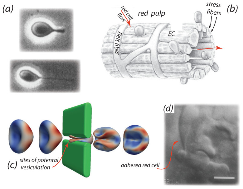
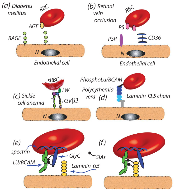
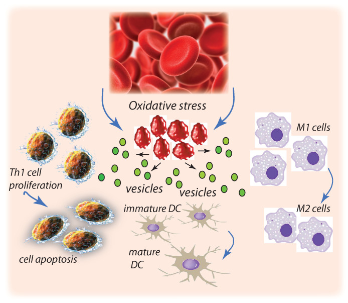
The red blood cell has become implicated in the progression of a range of diseases; mechanisms by which red cells are involved appear to include the transport of inflammatory species via red cell-derived vesicles. We review this role of RBCs in diseases such as diabetes mellitus, sickle cell anemia, polycythemia vera, central retinal vein occlusion, Gaucher disease, atherosclerosis, and myeloproliferative neoplasms. We propose a possibly unifying, and novel, paradigm for the inducement of RBC vesiculation during vascular flow of red cells adhered to the vascular endothelium as well as to the red pulp of the spleen. Indeed, we review the evidence for this hypothesis that links physiological conditions favoring both vesiculation and enhanced RBC adhesion and demonstrate the veracity of this hypothesis by way of a specific example occurring in splenic flow which we argue has various renderings in a wide range of vascular flows, in particular microvascular flows. We provide a mechanistic basis for membrane loss and the formation of lysed red blood cells in the spleen that may mediate their turnover. Our detailed explanation for this example also makes clear what features of red cell deformability are involved in the vesiculation process and hence require quantification and a new form of quantitative indexing.

Keywords: adhesion; hemolysis; vesiculation.


Conflict of interest statement

The authors declare no conflict of interest. There was no role of any funders in the writing or decision to publish this manuscript.

Figures

Figure 1
Figure 1
(a) Optical images of an RBC, adhered to a glass plate in a shear flow field forming an evagination and a tether (taken from Hochmuth et al. [72], with permission). (b) Schema of the splenic venous sinus and sinus wall taken from Drenckhahn and Wagner [94] (with permission). Note the stress fibers along the sides of intercellular slits whose caliber is mediated by the contractility of the myosin-actin filaments within these stress fibers [95,96,97]. Red blood cells are shown flowing through the slits. Note also the belt-like formations of the basement membrane ring fibers that abut the endothelium. Note also the “annular”, so marked in (b), or “ring”, fibers. (c) Influence of RBC initial orientation upon entering a venous splenic slit on the cell’s deformation shape. In this orientation the cell undergoes large deformations and develops an infolded region upon exiting the slit. (d) A snapshot of a red cell “hung up” while attempting passage thorough an IES; note this cell is not blocked but is adhered to the sinus endothelium in the IES (from MacDonald et al. [89], with permission).
Figure 2
Figure 2
SEM image taken from Fujita [111] showing transparent vesicles (white arrows) aggregated around a human splenic sinus (with permission). The size range of those vesicles visible was in the range 150–1000 nm.
Figure 3
Figure 3
(a,d) Side views of entire cell adhered to endothelial slit walls under shear stress of τ=0.072 Pa and τ=0.15 Pa, respectively; note encircled “tips” at the cell’s adhered zone. (b,e) Contours of skeletal areal deformation for the two levels of shear stress as in (a,d). (c,f) Show contours of contact stress (pressure) defined in the text and via (g) that is used as a guide. (h,i) Show two snapshots of skeletal areal deformation at the times indicated for the case of τ=0.072 Pa (γ˙=12 s1); note that (c) shows the snapshot of areal deformation at saturation for this case.
Figure 4
Figure 4
(a) Binding of Advanced Glycation End products (AGEs) of aged or disease RBCs to receptors for AGEs (RAGE) of the endothelium [37,131,132]. (b) Binding of exposed PS, due to membrane disruption or via activation of the phospholipid scramblase (PLSCR) to endothelial PS receptors (PSR) [27,107,123,135,137]. (c) Binding of integrins such as αvβ3 or αvβ1 to receptors such as adhesion molecule-4 (LW) on sickle cells, sRBC [33,34,35,138,139]. (d) Adherence of PV red cells to the endothelium by Lu/BCAM [19]. In (ad), N is the endothelial nucleus. (e,f) Loss of sialic acid residues due to erythrocyte aging allows sialic acid binding of Lu/BCAM to laminin-α5 sialic acid residues [42] in sickle cell anemia and PV.
Figure 5
Figure 5
Oxidatively damaged RBCs can augment mitogen-driven T-cell proliferation and apoptosis and Th1 proinflammatory, proatherogenic cytokine response. Oxidatively stressed RBCs that do not control LPS induced DC maturation promote DC maturation thereby inciting proinflammatory Th1 cell response. Oxidatively damaged, perhaps due to storage, may polarize macrophages toward M1 pathways also promoting proinflammatory cytokine response. In short, oxidative damage of RBCs promotes those cell phenotypes that promote vesiculation and can lead to atherosclerotic progression.

Similar articles

Cited by

References

    1. Buttari B., Profumo E., Riganò R. Crosstalk between Red Blood Cells and the Immune System and Its Impact on Atherosclerosis. BioMed Res. Int. 2015;2015:1–8. doi: 10.1155/2015/616834. - DOI - PMC - PubMed
    1. Boulanger C.M., Loyer X., Rautou P.-E., Amabile N. Extracellular vesicles in coronary artery disease. Nat. Rev. Cardiol. 2017;14:259–272. doi: 10.1038/nrcardio.2017.7. - DOI - PubMed
    1. Kim-Shapiro D.B., Lee J., Gladwin M.T. Storage lesion: Role of red blood cell breakdown. Transfusion. 2011;51:844–851. doi: 10.1111/j.1537-2995.2011.03100.x. - DOI - PMC - PubMed
    1. Businaro R., Tagliani A., Buttari B., Profumo E., Ippoliti F., Cristofano C.D., Capoano R., Salvati B., Riganò R. Cellular and molecular players in the atherosclerosis plaque progression. Ann. N. Y. Acad. Sci. 2012;1262:134–141. doi: 10.1111/j.1749-6632.2012.06600.x. - DOI - PubMed
    1. Jy W., Ricci M., Shariatmadar S., Gomez-Marin O., Horstman L.H., Ahn Y.S. Microparticles in stored red blood cells as potential mediators of transfusion complications. Transfusion. 2011;51:886–893. doi: 10.1111/j.1537-2995.2011.03099.x. - DOI - PMC - PubMed