. 2021 May 29;13(11):2679.
doi: 10.3390/cancers13112679.

Primary Gastrointestinal T/NK Cell Lymphoma

Affiliations

Primary Gastrointestinal T/NK Cell Lymphoma

Eun Kyung Kim et al. Cancers (Basel). .

Abstract

Primary gastrointestinal T/NK cell lymphoma (GI-TNKL) is an uncommon and heterogeneous group of lymphoid malignancies. We aimed to investigate their subtype distribution, clinicopathologic characteristics, and clinical outcomes. A total of 38 GI-TNKL cases and their clinical and pathological characteristics were analyzed. GI-TNKL occurred in adults with a median patient age in the sixth decade of life and showed a slight male predominance. The most common histologic type was extranodal NK/T-cell lymphoma, nasal type (ENKTL; 34.2%), followed by monomorphic epitheliotropic intestinal T-cell lymphoma (MEITL; 31.6%), intestinal T-cell lymphoma, NOS (ITCL, NOS, 18.4%), anaplastic large cell lymphoma, ALK-negative (ALCL, ALK-; 13.2%). The small intestine was the primary affected region. More than 90% of patients complained of various GI symptoms and cases with advanced Lugano stage, high IPI score, or bowel perforation that required emergent operation were not uncommon. GI-TNKL also showed aggressive behavior with short progression-free survival and overall survival. This thorough clinical and pathological descriptive analysis will be helpful for accurate understanding, diagnosis, and treatment.

Keywords: T/NK cell lymphoma; clinicopathologic features; gastrointestinal tract; intestinal lymphoma.


Conflict of interest statement

The authors declare no conflict of interest.

Figures
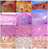
Figure 1
Figure 1
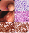
Primary gastrointestinal extranodal NK/T cell lymphoma, nasal type. Endoscopy reveals a huge ulceroinfiltrative lesion with an irregular base in the stomach (A). This case shows multiple hyperemic nodular lesions in the colon (B) and small bowel. The small bowel mucosa shows several various sized ulcerations (C). Microscopy shows diffuse transmural involvement (D) and a nodular mass in the low power view (E). Angiodestruction and necrosis are frequent findings (F). Tumor cells involve mucosa and submucosa and glands are relatively preserved (G). Medium to large atypical lymphoid cells infiltrate the lamina propria without epitheliotropism (H). Heterogeneous tumor cells show angiocentric pattern (I). Tumor cells are positive for granzyme B (J), CD56 (K), and EBV-encoded small RNA (L).
Figure 2
Figure 2
Monomorphic epitheliotropic intestinal T-cell lymphoma. The jejunum shows multiple ulceration with perforation (A). The transverse colon shows an ulceroinfiltrative mass involving mesenteric lymph nodes (B). Tumor cells are monotonous and medium-sized without inflammatory background (C). Some cases show variably admixed large pleomorphic cells (D). Tumor cells spread to the surrounding mucosa showing intraepithelial lymphocytosis (E) with relatively preserving villous architecture (F). Tumor cells are positive for CD8 (G), CD56 (H), and granzyme B (I).
Figure 3
Figure 3
Intestinal T-cell lymphoma, not otherwise specified. Stomach endoscopy shows multiple serpiginous-shaped superficial erosions with surface nodularity (A). Terminal ileum endoscopy demonstrates several geographic shallow and deep ulcerations (B). Tumor cells show diffuse and destructive growth (C). Some cases show medium to large atypical lymphoid cells with inflammatory background (D) or large pleomorphic cells with eosinophilic cytoplasm (F). Tumor cells are CD3 positive and reveal no epitheliotropism (D).
Figure 4
Figure 4
Anaplastic large cell lymphoma (ALCL). ALCL, ALK-negative (A,B). Endoscopically, the esophagus shows a large elevated submucosal tumor covered with mucosal ulceration (A). Biopsy findings demonstrate non-cohesive medium to large pleomorphic cells with irregular nuclei (B). ALCL, ALK-positive (CF). Endoscopy reveals hyperemic irregular and nodular surface with abnormalities of surrounding folds in the stomach (C). The same case shows large pleomorphic lymphoid cells with eccentric nuclei and abundant cytoplasm in the neutrophilic background (D). The tumor cells are immunoreactive for CD30 (cytoplasmic, nuclear membrane and Golgi region; E) and ALK (cytoplasm and nucleus; F).

Similar articles

Cited by

References

    1. Kohno S., Ohshima K., Yoneda S., Kodama T., Shirakusa T., Kikuchi M. Clinicopathological analysis of 143 primary malignant lymphomas in the small and large intestines based on the new WHO classification. Histopathology. 2003;43:135–143. doi: 10.1046/j.1365-2559.2003.01659.x. - DOI - PubMed
    1. Kim S.J., Choi C.W., Mun Y.-C., Oh S.Y., Kang H.J., Lee S.I., Won J.H., Kim M.K., Kwon J.H., Kim J.S., et al. Multicenter retrospective analysis of 581 patients with primary intestinal non-hodgkin lymphoma from the Consortium for Improving Survival of Lymphoma (CISL) BMC Cancer. 2011;11:321. doi: 10.1186/1471-2407-11-321. - DOI - PMC - PubMed
    1. Ding W., Zhao S., Wang J., Yang Q., Sun H., Yan J., Gao L., Yao W., Zhang W., Liu W. Gastrointestinal Lymphoma in Southwest China: Subtype Distribution of 1,010 Cases Using the WHO (2008) Classification in a Single Institution. Acta Haematol. 2016;135:21–28. doi: 10.1159/000437130. - DOI - PubMed
    1. Kim J.-M., Ko Y.-H., Lee S.-S., Huh J., Kang C.S., Kim C.W., Kang Y.K., Go J.H., Kim M.K., Kim W.-S., et al. WHO Classification of Malignant Lymphomas in Korea: Report of the Third Nationwide Study. Korean J. Pathol. 2011;45:254–260. doi: 10.4132/KoreanJPathol.2011.45.3.254. - DOI
    1. Swerdlow S.H., Campo E., Harris N.L., Jaffe E.S., Pileri S., Stein H., Thiele J., Swiatowa Organizacja Zdrowia. International Agency for Research on Cancer . WHO Classification of Tumours of Haematopoietic and Lymphoid Tissues. Revised 4th ed. International Agency for Research on Cancer; Lyon, France: 2017.

LinkOut - more resources