Enhanced Cellular Uptake in an Electrostatically Interacting Fucoidan-L-Arginine Fiber Complex
- PMID: 34072354
- PMCID: PMC8198147
- DOI: 10.3390/polym13111795
Enhanced Cellular Uptake in an Electrostatically Interacting Fucoidan-L-Arginine Fiber Complex
Abstract
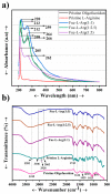
Fucoidan is an abundant marine sulfated polysaccharide extracted from the cell wall of brown macroalgae (seaweed). Recently, fucoidan has been highly involved in various industrial applications, such as pharmaceuticals, biomedicals, cosmetics, and food. However, the presence of a sulfate group (negative surface charge) in the fucoidan structure limits its potential and biological activity for use in biomedical applications during cellular uptake. Thus, we aimed to improve the uptake of fucoidan by using an L-arginine uptake enhancer within an in vitro study. A Fucoidan-L-Arginine (Fuc-L-Arg) fiber complex was prepared via α-helical electrostatic interactions using a freeze-drying technique and confirmed using field-emission scanning electron microscopy, Fourier transform infrared spectroscopy, and nuclear magnetic resonance spectroscopy. In addition, fucoidan was conjugated with cyanine 3 (Cy3) dye to track its cellular uptake. Furthermore, the results of Fuc-L-Arg (1:1, 1:2.5) complexes revealed biocompatibility >80% at various concentrations (5, 10, 25, 50, 100 µg/mL). Owing to the higher internalization of the Fuc-L-Arg (1:5) complex, it exhibited <80% biocompatibility at higher concentrations (25, 50, 100 µg/mL) of the complex. In addition, improved cellular internalization of Fuc-L-Arg complexes (1:5) in HeLa cells have been proved via flow cytometry quantitative analysis. Hence, we highlight that the Fuc-L-Arg (1:5) fiber complex can act as an excellent biocomplex to exhibit potential bioactivities, such as targeting cancers, as fucoidan shows higher permeability in HeLa cells.
Keywords: L-arginine; cyanine 3 dye; fucoidan.
Conflict of interest statement
The authors declare no conflict of interest.
Figures


Similar articles
-
Structures of oligosaccharides derived from Cladosiphon okamuranus fucoidan by digestion with marine bacterial enzymes.Mar Biotechnol (NY). 2003 Nov-Dec;5(6):536-44. doi: 10.1007/s10126-002-0107-9. Epub 2003 Jul 17. Mar Biotechnol (NY). 2003. PMID: 14730423
-
Nano-Sized Fucoidan Interpolyelectrolyte Complexes: Recent Advances in Design and Prospects for Biomedical Applications.Int J Mol Sci. 2023 Jan 30;24(3):2615. doi: 10.3390/ijms24032615. Int J Mol Sci. 2023. PMID: 36768936 Free PMC article. Review.
-
Structure and rheological characteristics of fucoidan from sea cucumber Apostichopus japonicus.Food Chem. 2015 Aug 1;180:71-76. doi: 10.1016/j.foodchem.2015.02.034. Epub 2015 Feb 14. Food Chem. 2015. PMID: 25766803
-
Structural characterization and effect on leukopenia of fucoidan from Durvillaea antarctica.Carbohydr Polym. 2021 Mar 15;256:117529. doi: 10.1016/j.carbpol.2020.117529. Epub 2020 Dec 17. Carbohydr Polym. 2021. PMID: 33483047
-
Fucoidan, a brown seaweed polysaccharide in nanodrug delivery.Drug Deliv Transl Res. 2023 Oct;13(10):2427-2446. doi: 10.1007/s13346-023-01329-4. Epub 2023 Apr 3. Drug Deliv Transl Res. 2023. PMID: 37010790 Review.
Cited by
-
Characterization and Bioactivity of Polysaccharides Separated through a (Sequential) Biorefinery Process from Fucus spiralis Brown Macroalgae.Polymers (Basel). 2022 Sep 30;14(19):4106. doi: 10.3390/polym14194106. Polymers (Basel). 2022. PMID: 36236054 Free PMC article.
References
-
- Pacheco D., García-Poza S., Cotas J., Gonçalves A.M., Pereira L. Fucoidan—A valuable source from the ocean to pharmaceutical. Front. Drug Chem. Clin. Res. 2020;3:1–4.
Grants and funding
- MOST 105-2221-E-011- 151-MY3 and 105-2221-E-011-133-MY3/Ministry of Science and Technology of the Republic of China (Taiwan)
- (Grant Nos. TMU-NTUST-104-10 and TMUNTUST-105-07)/National Taiwan University of Science and Technology, National Taiwan university and Taipei Medical University Joint Research Program
LinkOut - more resources
Full Text Sources
Other Literature Sources

