Review
. 2021 May 29;13(6):1025.
doi: 10.3390/v13061025.

Insights into SARS-CoV-2 Persistence and Its Relevance

Affiliations
Review

Insights into SARS-CoV-2 Persistence and Its Relevance

Belete A Desimmie et al. Viruses. .

Abstract

Severe acute respiratory syndrome coronavirus 2 (SARS-CoV-2), the causative agent of coronavirus disease 2019 (COVID-19), continues to wreak havoc, threatening the public health services and imposing economic collapse worldwide. Tailoring public health responses to the SARS-CoV-2 pandemic depends on understanding the mechanism of viral replication, disease pathogenesis, accurately identifying acute infections, and mapping the spreading risk of hotspots across the globe. However, effective identification and isolation of persons with asymptomatic and mild SARS-CoV-2 infections remain the major obstacles to efforts in controlling the SARS-CoV-2 spread and hence the pandemic. Understanding the mechanism of persistent viral shedding, reinfection, and the post-acute sequalae of SARS-CoV-2 infection (PASC) is crucial in our efforts to combat the pandemic and provide better care and rehabilitation to survivors. Here, we present a living literature review (January 2020 through 15 March 2021) on SARS-CoV-2 viral persistence, reinfection, and PASC. We also highlight potential areas of research to uncover putative links between viral persistence, intra-host evolution, host immune status, and protective immunity to guide and direct future basic science and clinical research priorities.

Keywords: COVID-19; PASC; SARS-CoV-2; coronaviruses; long COVID; reinfection; viral persistence.


Conflict of interest statement

The authors declare no conflict of interest.

Figures
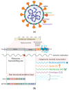
Figure 1
Figure 1
SARS-CoV-2 genome organization and replication cycle. (a) The SARS-CoV-2 virion structure. While the viral membrane and envelope proteins ensure its genomic RNA incorporation and assembly, the trimeric spike (S) protein provides specificity and high-affinity binding for its receptor to enter into target cells. The positive-sense, single-stranded RNA genome (+ssRNA) is encapsidated by the nucleocapsid. (b) Schematic depiction of SARS-CoV-2 genome architecture and the poly-(proteins) it encodes. (c) Schematics of SARS-CoV-2 replication: (1) SARS-CoV-2 virions bind to ACE2, its cellular receptor, and the type 2 transmembrane serine protease (TMPRSS2), a host factor that promotes viral particles entry and fusion at the plasma membrane or endosomes. (2) In the cytosol, the incoming gRNA will be released and subjected to immediate translation of the ORF1a and ORF1b open reading frame resulting in the polyproteins (pp1a and pp1ab), which are further proteolytically processed to the components of the replicase complex. (3) The replicase complex is an assemblage of multiple nonstructural proteins (nsps) that orchestrate transcription of the viral RNAs and viral replication. Concordantly, the virus compels the host cell to create the viral replication organelles, including the perinuclear double-membrane vesicles (DMV), the convoluted membranes, and small, open double-membrane spherules (DMS). These structures provide a protective microenvironment for the viral gRNA and subgenomic mRNA during replication and transcription. (4,5) The newly synthesized surface structural proteins translocate to the ER membrane and transit through the ER-to-Golgi intermediate compartment (ERGIC), which then assembles and encapsidates the ribonucleocapsid. (6,7) Ultimately, the progeny virions bud into the lumen of the secretory vesicles and will be released by exocytosis.
Figure 1
Figure 1
SARS-CoV-2 genome organization and replication cycle. (a) The SARS-CoV-2 virion structure. While the viral membrane and envelope proteins ensure its genomic RNA incorporation and assembly, the trimeric spike (S) protein provides specificity and high-affinity binding for its receptor to enter into target cells. The positive-sense, single-stranded RNA genome (+ssRNA) is encapsidated by the nucleocapsid. (b) Schematic depiction of SARS-CoV-2 genome architecture and the poly-(proteins) it encodes. (c) Schematics of SARS-CoV-2 replication: (1) SARS-CoV-2 virions bind to ACE2, its cellular receptor, and the type 2 transmembrane serine protease (TMPRSS2), a host factor that promotes viral particles entry and fusion at the plasma membrane or endosomes. (2) In the cytosol, the incoming gRNA will be released and subjected to immediate translation of the ORF1a and ORF1b open reading frame resulting in the polyproteins (pp1a and pp1ab), which are further proteolytically processed to the components of the replicase complex. (3) The replicase complex is an assemblage of multiple nonstructural proteins (nsps) that orchestrate transcription of the viral RNAs and viral replication. Concordantly, the virus compels the host cell to create the viral replication organelles, including the perinuclear double-membrane vesicles (DMV), the convoluted membranes, and small, open double-membrane spherules (DMS). These structures provide a protective microenvironment for the viral gRNA and subgenomic mRNA during replication and transcription. (4,5) The newly synthesized surface structural proteins translocate to the ER membrane and transit through the ER-to-Golgi intermediate compartment (ERGIC), which then assembles and encapsidates the ribonucleocapsid. (6,7) Ultimately, the progeny virions bud into the lumen of the secretory vesicles and will be released by exocytosis.
Figure 2
Figure 2
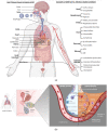
SARS-CoV-2 tropism, clinical presentation, and pathophysiology of alveolar damage. (a) A wide range of clinical and laboratory abnormalities can be observed in SARS-CoV-2 infection. In severe COVID-19 cases, the pathophysiology of the disease involves accelerated viral replication, cytokine storm, hyperinflammatory state, systemic endotheliitis, and hypercoagulability with secondary organ dysfunction (often pulmonary, cardiovascular, renal, or hepatic). (b) When SARS-CoV-2 makes it to the lower airway and infects type II pneumocytes (right side), the accelerated replication of the virus induces host cell death by pyroptosis and release of proinflammatory and damage-associated molecules, further activating and recruiting proinflammatory immune cells, including neutrophils, lymphocytes, and monocytes to the site of infection. In a defective or misfiring immune response, there will be an overproduction of proinflammatory and profibrotic cytokines, resulting in diffuse alveolar damage with fibrin-rich hyaline membrane deposition and loss of the gas exchange function of the alveoli, and hence development of hypoxic respiratory failure. Furthermore, the resultant cytokine storm leads to multi-organ damage. However, in a healthy immune response, virus-specific immune cells will be recruited and eliminate the infected cells before the virus spreads to ultimately minimize the alveolar damage (the left side). AMS denotes altered mental status, ARDS denotes acute respiratory distress syndrome, AKI denotes acute kidney injury, and ATN denotes acute tubular necrosis.
Figure 3
Figure 3
Time course of SARS-CoV-2 infection, clinical spectrum, and immune response. (a) Kinetics of viral load and clinical spectrum. (b) A schematic representation of immune response following SARS-CoV-2 exposure. The innate immune response appears shortly after exposure (purple line) accompanied by cytokine storm (purple dashed line), followed by adaptive immunity development after the first week post-infection. Seroconversion and T cell response occur during the second week after symptoms onset. While IgM peaks early and decays faster, IgG titers and T cell response detected around day 10 and their peak and height are likely to be influenced by disease severity and virus load. The level of antibody protection from reinfection, the duration of the total humoral immune response above this protective threshold (dashed black horizontal line), and the rate of decline from mild- or severe-infection-induced antibodies is not known for SARS-CoV-2 during the PASC period.
Figure 4
Figure 4
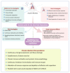
Outcomes of SARS-CoV-2 infection. During the acute infection, the host and the virus compete for dominance to tip the balance in their favor, resulting in different outcomes as indicated in the highlighted boxes. If the host strategies prevail to achieve complete recovery, the virus will be cleared. Another outcome that is poorly understood and yet getting traction is the multisystemic sequelae of SARS-CoV-2 infection called PASC. On the other hand, if the virus wins the battle, a variety of outcomes are expected, as indicated in the illustration. Viral persistence in a minority of patients, particularly in immunocompromised patients, has now been well documented; however, the underlying mechanism is unknown. There is a possibility of establishing metastable equilibrium between the host and SARS-CoV-2 to facilitate persistence and/or chronic infection.

Similar articles

Cited by

References

    1. WHO Coronavirus Disease (COVID-19) Pandemic. [(accessed on 19 April 2021)]; Available online: https://www.who.int/emergencies/diseases/novel-coronavirus-2019.
    1. Coronaviridae Study Group of the International Committee on Taxonomy of Viruses The species Severe acute respiratory syndrome-related coronavirus: Classifying 2019-nCoV and naming it SARS-CoV-2. Nat. Microbiol. 2020;5:536–544. doi: 10.1038/s41564-020-0695-z. - DOI - PMC - PubMed
    1. Zhou P., Yang X.L., Wang X.G., Hu B., Zhang L., Zhang W., Si H.R., Zhu Y., Li B., Huang C.L., et al. A pneumonia outbreak associated with a new coronavirus of probable bat origin. Nature. 2020;579:270–273. doi: 10.1038/s41586-020-2012-7. - DOI - PMC - PubMed
    1. Zhu N., Zhang D., Wang W., Li X., Yang B., Song J., Zhao X., Huang B., Shi W., Lu R., et al. A Novel Coronavirus from Patients with Pneumonia in China, 2019. N. Engl. J. Med. 2020;382:727–733. doi: 10.1056/NEJMoa2001017. - DOI - PMC - PubMed
    1. Wu F., Zhao S., Yu B., Chen Y.M., Wang W., Song Z.G., Hu Y., Tao Z.W., Tian J.H., Pei Y.Y., et al. A new coronavirus associated with human respiratory disease in China. Nature. 2020;579:265–269. doi: 10.1038/s41586-020-2008-3. - DOI - PMC - PubMed

Publication types