Review
. 2021 May 29;10(11):2412.
doi: 10.3390/jcm10112412.

Metabolic Alterations in Sepsis

Affiliations
Review

Metabolic Alterations in Sepsis

Weronika Wasyluk et al. J Clin Med. .

Abstract

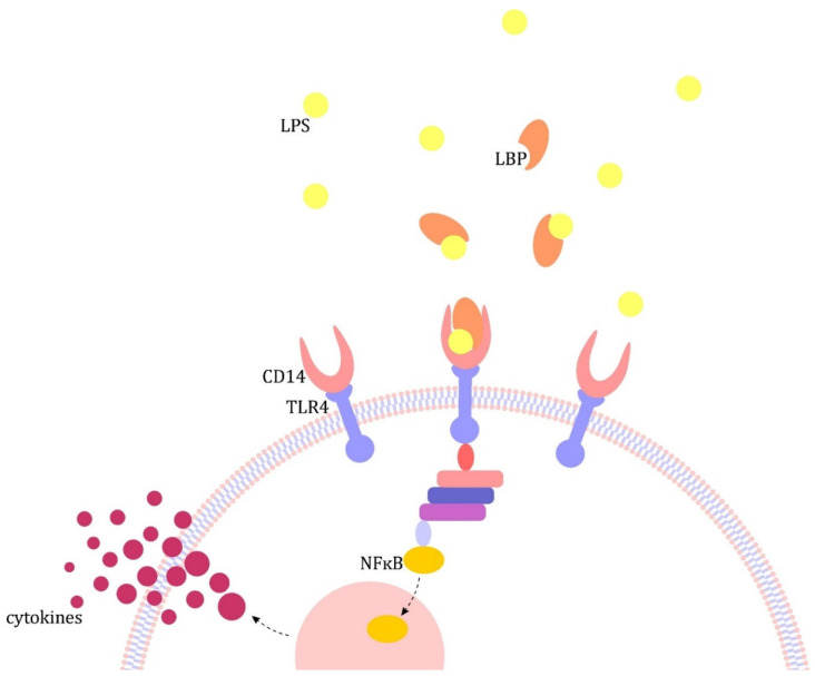
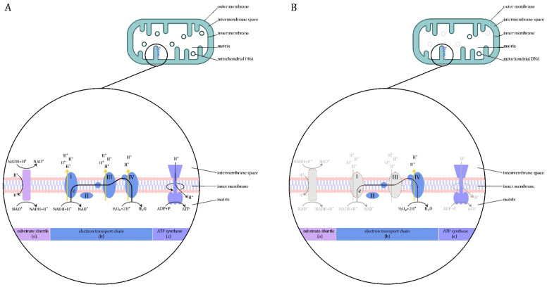
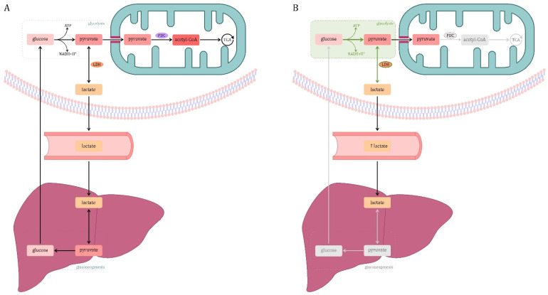
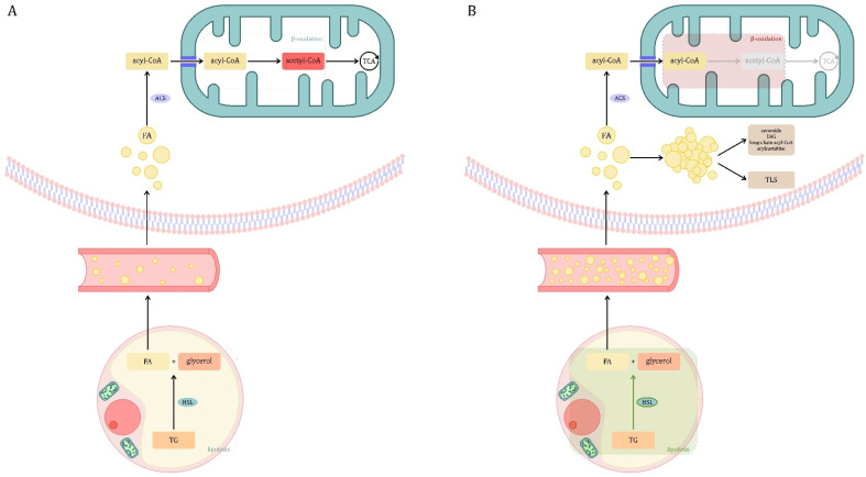
Sepsis is defined as "life-threatening organ dysfunction caused by a dysregulated host response to infection". Contrary to the older definitions, the current one not only focuses on inflammation, but points to systemic disturbances in homeostasis, including metabolism. Sepsis leads to sepsis-induced dysfunction and mitochondrial damage, which is suggested as a major cause of cell metabolism disorders in these patients. The changes affect the metabolism of all macronutrients. The metabolism of all macronutrients is altered. A characteristic change in carbohydrate metabolism is the intensification of glycolysis, which in combination with the failure of entering pyruvate to the tricarboxylic acid cycle increases the formation of lactate. Sepsis also affects lipid metabolism-lipolysis in adipose tissue is upregulated, which leads to an increase in the level of fatty acids and triglycerides in the blood. At the same time, their use is disturbed, which may result in the accumulation of lipids and their toxic metabolites. Changes in the metabolism of ketone bodies and amino acids have also been described. Metabolic disorders in sepsis are an important area of research, both for their potential role as a target for future therapies (metabolic resuscitation) and for optimizing the current treatment, such as clinical nutrition.

Keywords: critical illness; intensive care; metabolic disorders; metabolism; mitochondria; sepsis; septic shock.


Conflict of interest statement

The authors declare no conflict of interest.

Figures

Figure 1
Figure 1
Pathogenesis of sepsis on the example of lipopolysaccharide (LPS) [4]. The major pathomechanism is the formation of complexes in the blood with lipopolysaccharide binding protein (LBP), which in turn binds to CD14 receptors present on monocytes/macrophages and neutrophils and circulating in the plasma. The resulting complex activates Toll-like receptors (TLRs), which transmit the signal inside the cell, leading to the translocation of the nuclear factor kB (NF-kB) to the cell nucleus and activation of pro-inflammatory cytokine gene promoters, including interleukin (IL) 1 and tumor necrosis factor α (TNF-α) [9].
Figure 2
Figure 2
Production of energy in the mitochondria. (A) Physiological conditions. Mitochondria are formed from the permeable outer membrane, the intermembrane space, the selectively permeable inner membrane and the matrix. In the inner membrane, there are electron transport chain enzymes and ATP synthase. The tricarboxylic acid (TCA) cycle and β-oxidation enzymes are contained in the mitochondrion. The energy released during the oxidation of energy substrates is available inside the mitochondria in the form of reducing equivalents. The electron transport chain (ETC) (b) collects and transports the reducing equivalents, directing them to react with oxygen to form water. ETC is made up of four large protein complexes embedded in the inner mitochondrial membrane. The flow of electrons through the respiratory chain is driven by the redox potential difference and is mediated by three of the complexes (I, III, IV), substrates having a more positive potential than NAD+/NADH transfer electrons to complex III via complex II (instead of I). The flow of electrons through complexes I, III and IV causes the displacement of protons through the inner mitochondrial membrane—from the matrix to the intermembrane space. This allows the creation of a proton gradient that drives the synthesis of ATP. Proton-motive force (PMF), created by ions accumulating in the mesothelial space, is used by the ATP synthase (c) located in the inner mitochondrial membrane—it attaches a phosphate residue to adenosine diphosphate, creating ATP [31]. The inner mitochondrial membrane is impermeable to nicotinamide adenine dinucleotide (NADH), which is continuously produced in the cytosol by the glycolytic pathway. The transfer of reducing equivalents from NADH across the inner mitochondrial membrane requires the presence of a pair of dehydrogenase-conjugated substrates on both sides of the membrane, a system known as a substrate shuttles. Two types of substrate shuttles are involved in the transfer of reducing equivalents across the inner mitochondrial membrane—the glycerophosphate shuttle and the malate-aspartate shuttle (a) [31]. (B) Sepsis. In the course of sepsis, the expression and activity of some electron transport chain (ETC) complexes (b) are reduced. This may reduce the efficiency of ETC, whose role is to generate a proton gradient that drives the synthesis of ATP (c). Loss of some copies of mitochondrial DNA (mtDNA), which contains information on key subunits of the ECT complexes, has also been reported. In addition, sepsis can impair the operation of the substrate shuttle. Legend: gray color—insufficient ETC complexes and disturbed cellular processes.
Figure 3
Figure 3
Glucose metabolism. (A) Physiological conditions. Glycolysis is the conversion of glucose into pyruvate. In some cases, for example, when glycolysis occurs anaerobically or in cells incapable of pyruvate oxidation, pyruvate may be converted to lactate by the enzyme lactate dehydrogenase (LDH). Blood lactate (mainly formed in skeletal muscle) is taken up by the liver, converted to pyruvate and incorporated into the gluconeogenesis pathway (Cori cycle). In other cases, the pyruvate formed in the cytosol is transported to the mitochondria by a proton symporter. In the mitochondrion, pyruvate undergoes oxidative decarboxylation to acetyl-CoA mediated by the multi-enzyme pyruvate dehydrogenase complex (PDC). Oxidation of pyruvate to acetyl-CoA connects glycolysis with the tricarboxylic acid (TCA) cycle [74]. (B) Sepsis. This is due, inter alia, to a decrease in pyruvate dehydrogenase complex (PDC) activity. One explanation is the phosphorylation of the enzyme by pyruvate dehydrogenase kinases (PDKs) and its decreased expression in sepsis. Moreover, in sepsis, the hypoxia-inducible factor 1α (HIF-1α) is activated, which is one of the main mediators of glycolysis. HIF-1α regulates the expression of genes of enzymes related to glycolysis (hexokinase, phosphofructokinase-1, glucose-6-phosphate dehydrogenase, lactate dehydrogenase, pyruvate dehydrogenase kinase, glutamate transporter-1). The intensification of glycolysis in combination with the failure of entering pyruvate to the tricarboxylic acid (TCA) cycle increases the formation of lactate. A decrease in hepatic lactate-based gluconeogenesis has been reported. This increases the production of lactate, while blocking the main routes of its disposal, which results in an increase in its concentration in the blood. Legend: gray color—inefficient cellular processes; green color—increasing the activity of the process or enzyme.
Figure 4
Figure 4
Lipid metabolism. (A) Physiological conditions. Triglycerides (TG) stored in adipocytes must undergo a three-step process by which other tissues can access their stored energy reserves. The first step, lipid mobilization, involves lipolysis of TG to fatty acids (FA) and glycerol, which diffuse into the plasma and are transported to the target tissues. TG is lipolyzed by the enzyme hormone-sensitive lipase (HSL). This process is influenced by many hormones. Insulin inhibits the action of HSL. Lipolysis-promoting hormones include catecholamines, glucagon, adrenocorticotropic hormone (ACTH), thyroid-stimulating hormone (TSH), growth hormone (GH) and vasopressin. Moreover, most lipolytic processes require the presence of thyroid hormones and glucocorticosteroids for optimal effect [114]. The second step is to activate FA and transport them to the mitochondria. The enzyme acyl-CoA synthase catalyzes the conversion of FA to active fatty acid (acyl-CoA). Then, long-chain acyl-CoA molecules penetrate the inner mitochondrial membrane as carnitine derivatives (acylcarnitine). The third step is β-oxidation—acyl-CoA molecules are broken down into acetyl-CoA, which can be oxidized in the tricarboxylic acid (TCA) cycle [115]. (B) Sepsis. In the initial stage of sepsis, lipolysis in adipose tissue is upregulated. The role of hormone-sensitive lipase (HSL) activation under the influence of LPS and prolipolytic hormones is suggested. Insulin resistance also contributes to the activation of lipolysis. Patients with sepsis have increased levels of fatty acids (FA) and triglycerides (TG) in the blood. This may be due to insulin resistance in the liver and adipose tissue. In the course of sepsis, the β-oxidation process is disturbed, which may be associated with a decrease in the expression of genes belonging to the PPAR-α signaling pathway. Additionally, the transport of long-chain FA to the mitochondria may be impaired due to possible accumulation of malonyl-CoA generated from glucose in hepatocytes. The lower efficiency of β-oxidation can cause energy deficiency, lipotoxicity and mitochondrial dysfunction, leading to organ damage. Lipotoxicity is a pathological metabolic phenomenon resulting from the accumulation of lipid intermediates in tissue other than adipose tissue. Lipid accumulation may lead to some of their metabolites (e.g., diacylglycerol (DAG), ceramide) reaching levels potentially harmful to cells. It is believed that excess lipids can be diverted to non-oxidative pathways and be transformed into toxic lipid species (TLS), which can damage mitochondria, modify cell signaling and increase apoptosis (lipoapoptosis). Legend: gray color—inefficient cellular processes; green color—increasing the activity of the process or enzyme; red color—lowering the activity of the process or enzyme.

Similar articles

Cited by

References

    1. Singer M., Deutschman C.S., Seymour C.W., Shankar-Hari M., Annane D., Bauer M., Bellomo R., Bernard G.R., Chiche J.D., Coopersmith C.M., et al. The third international consensus definitions for sepsis and septic shock (sepsis-3) JAMA. 2016;315:801–810. doi: 10.1001/jama.2016.0287. - DOI - PMC - PubMed
    1. Cohen J., Vincent J.L., Adhikari N.K., Machado F.R., Angus D.C., Calandra T., Jaton K., Giulieri S., Delaloye J., Opal S., et al. Sepsis: A roadmap for future research. Lancet Infect. Dis. 2015;15:581–614. doi: 10.1016/S1473-3099(15)70112-X. - DOI - PubMed
    1. Kenig A., Ilan Y. A Personalized Signature and Chronotherapy-Based Platform for Improving the Efficacy of Sepsis Treatment. Front. Physiol. 2019;10:1542. doi: 10.3389/fphys.2019.01542. - DOI - PMC - PubMed
    1. Wasyluk W., Wasyluk M., Zwolak A. Sepsis as a Pan-Endocrine Illness—Endocrine Disorders in Septic Patients. J. Clin. Med. 2021;10:2075. doi: 10.3390/jcm10102075. - DOI - PMC - PubMed
    1. Carré J., Singer M., Moncada S. Nitric Oxide. In: Abraham E., Singer M., editors. Mechanisms of Sepsis-Induced Organ Dysfunction and Recovery. Springer; Berlin/Heidelberg, Germany: New York, NY, USA: 2007. pp. 77–95.

LinkOut - more resources