. 2021 May 29;12(6):838.
doi: 10.3390/genes12060838.

Response of Bovine Cumulus-Oocytes Complexes to Energy Pathway Inhibition during In Vitro Maturation

Affiliations

Response of Bovine Cumulus-Oocytes Complexes to Energy Pathway Inhibition during In Vitro Maturation

Paulina Lipinska et al. Genes (Basel). .

Abstract

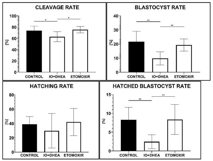
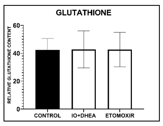
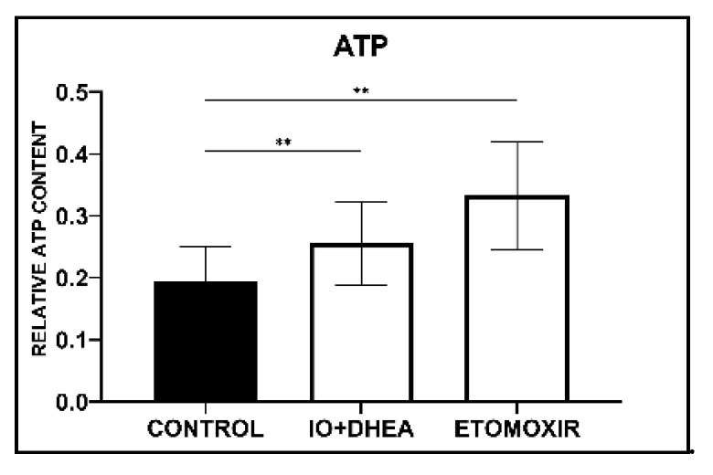
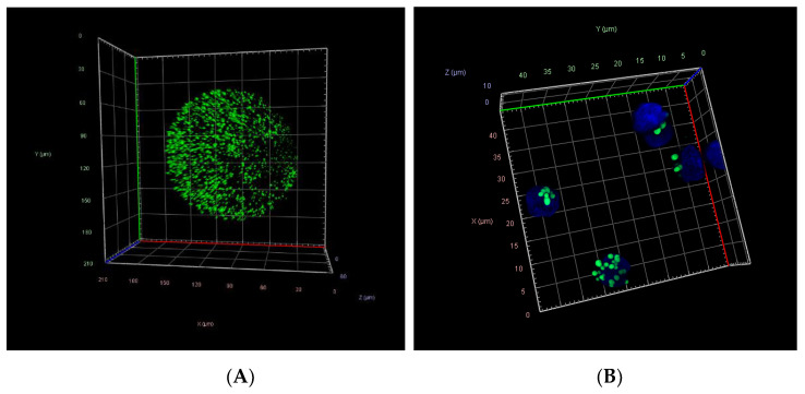
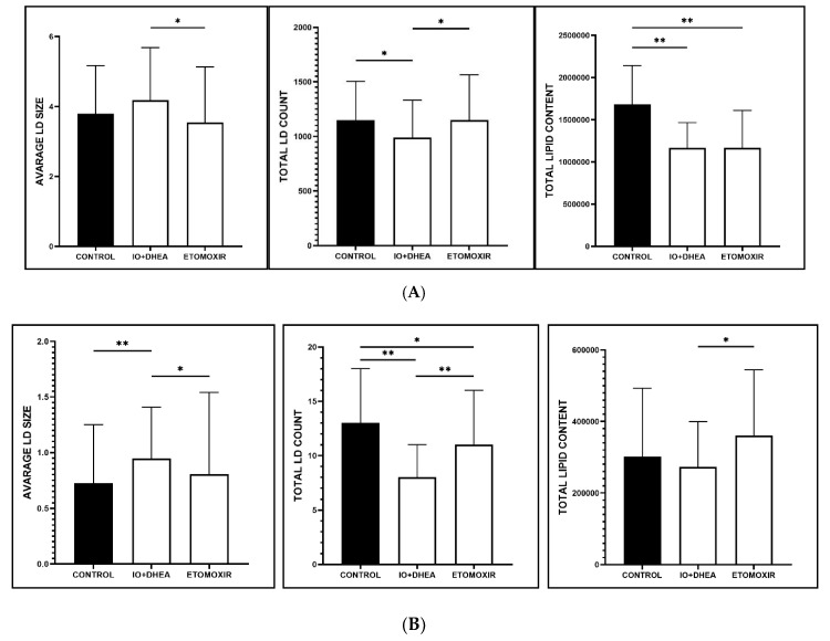
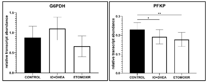
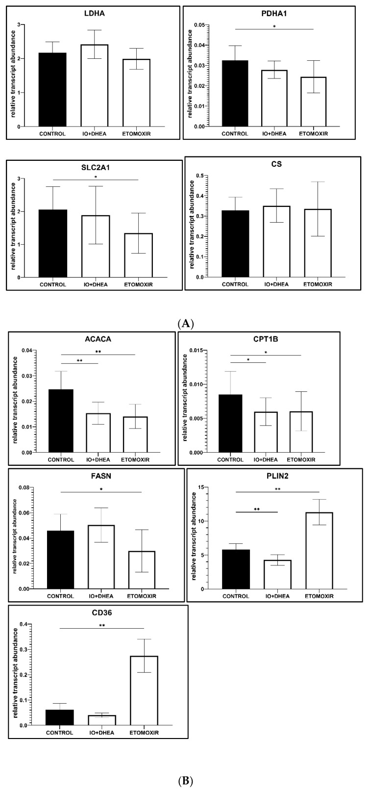
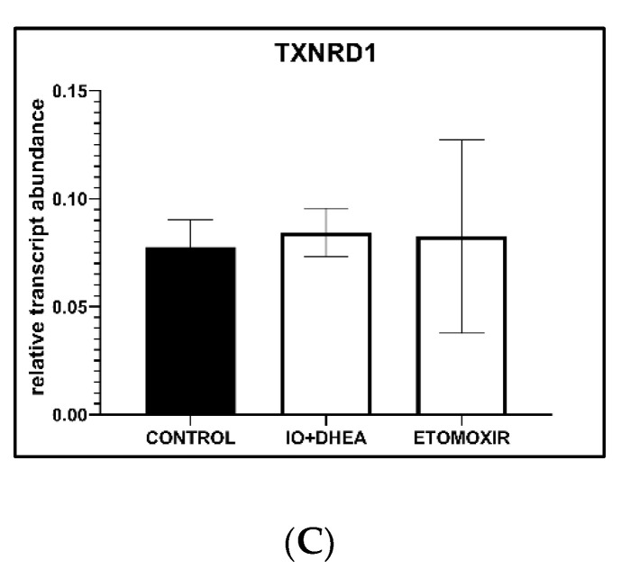
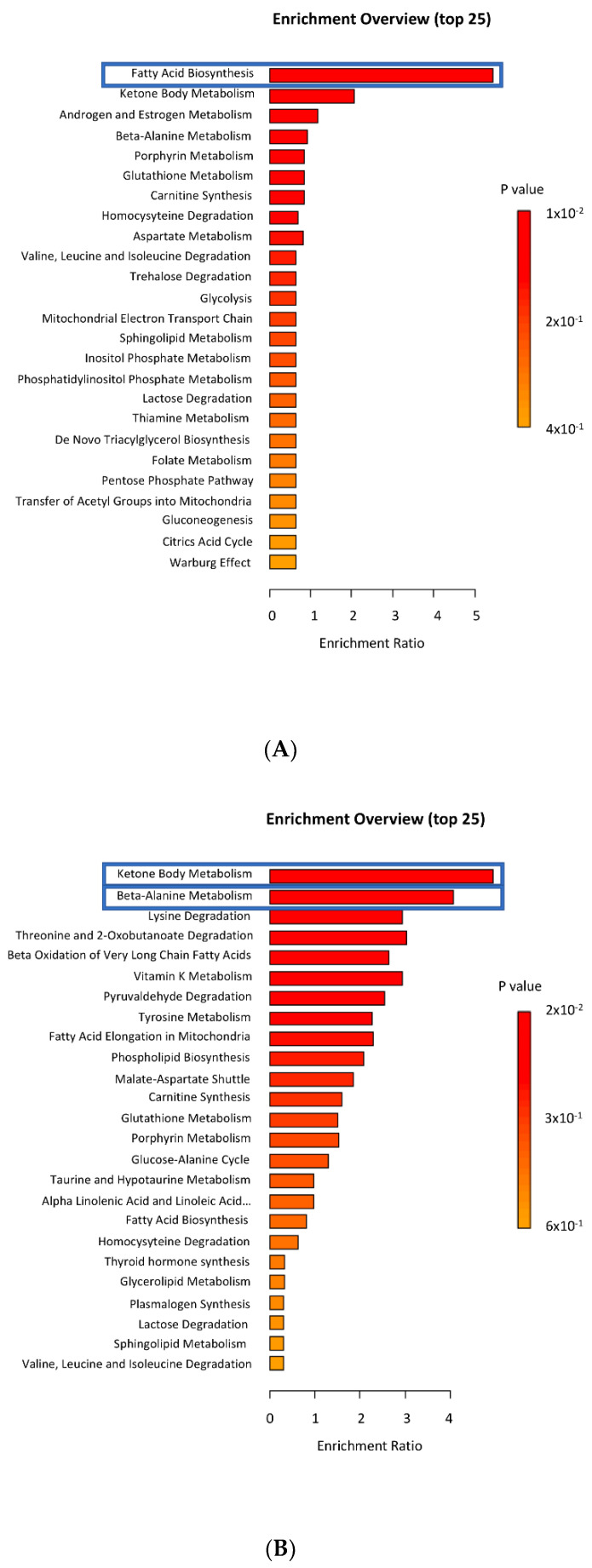
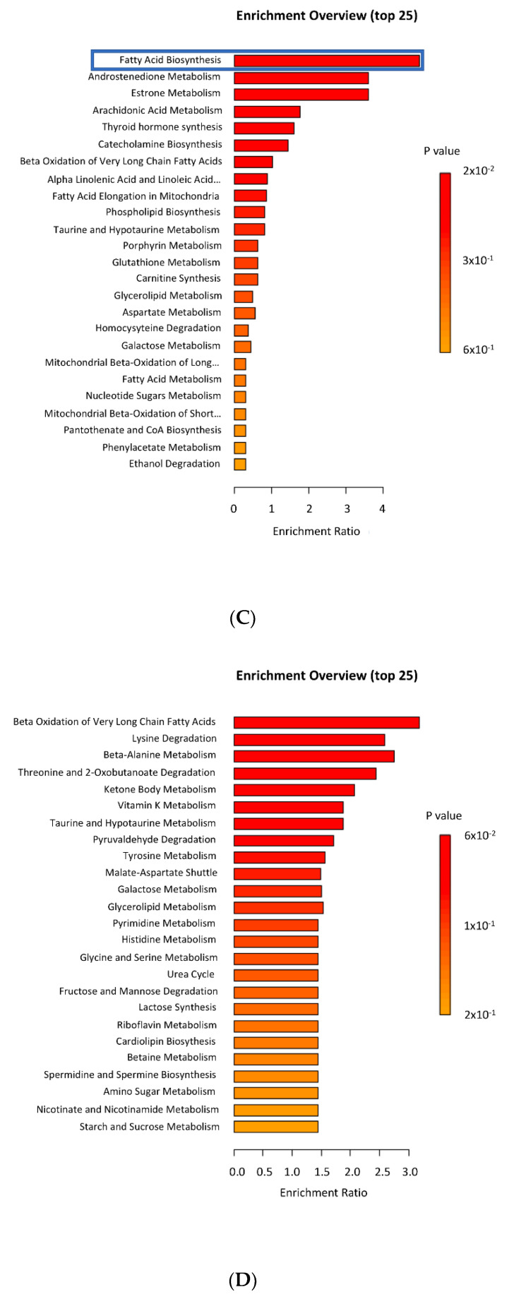
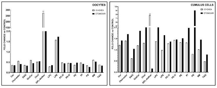
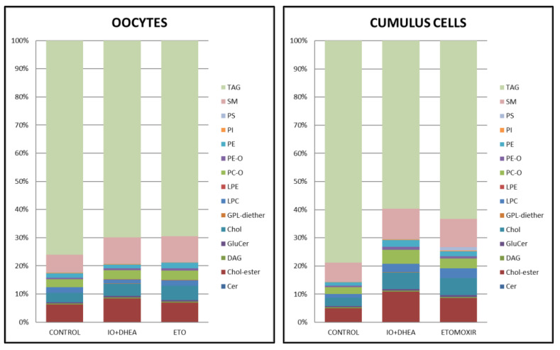
Glucose or fatty acids (FAs) metabolisms may alter the ovarian follicle environment and thus determine oocyte and the nascent embryo quality. The aim of the experiment was to investigate the effect of selective inhibition of glucose (iodoacetate + DHEA) or FA (etomoxir) metabolism on in vitro maturation (IVM) of bovine COCs (cumulus-oocyte complexes) to investigate oocyte's development, quality, and energy metabolism. After in vitro fertilization, embryos were cultured to the blastocyst stage. Lipid droplets, metabolome, and lipidome were analyzed in oocytes and cumulus cells. mRNA expression of the selected genes was measured in the cumulus cells. ATP and glutathione relative levels were measured in oocytes. Changes in FA content in the maturation medium were evaluated by mass spectrometry. Our results indicate that only glucose metabolism is substantial to the oocyte during IVM since only glucose inhibition decreased embryo culture efficiency. The most noteworthy differences in the reaction to the applied inhibition systems were observed in cumulus cells. The upregulation of ketone body metabolism in the cumulus cells of the glucose inhibition group suggest possibly failed attempts of cells to switch into lipid consumption. On the contrary, etomoxir treatment of the oocytes did not affect embryo development, probably due to undisturbed metabolism in cumulus cells. Therefore, we suggest that the energy pathways analyzed in this experiment are not interchangeable alternatives in bovine COCs.

Keywords: cumulus cells; energy metabolism; fatty acids; glucose; oocyte.


Conflict of interest statement

The authors declare no conflict of interest.

Figures

Figure 1
Figure 1
The effect of glucose (IO+DHEA) and fatty acid (ETOMOXIR) metabolism inhibition on the efficiency of in vitro embryo production (means ± SD). *—p ≤ 0.05, **—p ≤ 0.01.
Figure 2
Figure 2
The effect of glucose (IO+DHEA) and fatty acid (ETOMOXIR) metabolism inhibition on glutathione content in mature oocytes (means ± SD).
Figure 3
Figure 3
The effect of glucose (IO+DHEA) and fatty acid (ETOMOXIR) metabolism inhibition on ATP content in oocytes (means ± SD). **—p ≤ 0.01.
Figure 4
Figure 4
Confocal 3D projections of fluorescently stained oocytes (A) and cumulus cells (B) with BODIPY 493/503 (green—lipid droplets) and 4′,6-diamidino-2-phenylindole (DAPI; blue—nuclei).
Figure 5
Figure 5
The effect of glucose (IO+DHEA) and fatty acid (ETOMOXIR) metabolism inhibition on the lipid droplets parameters in (A) oocytes and (B) cumulus cells (means ± SD). *—p ≤ 0.05, **—p ≤ 0.01.
Figure 6
Figure 6
The effect of glucose (IO+DHEA) and fatty acid (ETOMOXIR) metabolism inhibition on the selected gene expression levels (mRNA) in the cumulus cells. The results are shown as a transcript abundance relative to the geometric mean of reference genes (means ± SEM). *—p ≤ 0.05, **—p ≤ 0.01: (A) genes involved in glucose metabolism; (B) genes involved in fatty acid metabolism; (C) genes involved in oxidative stress protection.
Figure 6
Figure 6
The effect of glucose (IO+DHEA) and fatty acid (ETOMOXIR) metabolism inhibition on the selected gene expression levels (mRNA) in the cumulus cells. The results are shown as a transcript abundance relative to the geometric mean of reference genes (means ± SEM). *—p ≤ 0.05, **—p ≤ 0.01: (A) genes involved in glucose metabolism; (B) genes involved in fatty acid metabolism; (C) genes involved in oxidative stress protection.
Figure 7
Figure 7
Enrichment metabolites analysis in oocytes and cumulus cells matured with IO+DHEA or ETOMOXIR inhibitors (data compared to control). Pathways that differed significantly (p ≤ 0.05) were marked with blue squares: (A) control vs. IO+DHEA in oocytes; (B) control vs. IO+DHEA in cumulus cells; (C) control vs. ETOMOXIR in oocytes; (D) control vs. ETOMOXIR in cumulus cells.
Figure 7
Figure 7
Enrichment metabolites analysis in oocytes and cumulus cells matured with IO+DHEA or ETOMOXIR inhibitors (data compared to control). Pathways that differed significantly (p ≤ 0.05) were marked with blue squares: (A) control vs. IO+DHEA in oocytes; (B) control vs. IO+DHEA in cumulus cells; (C) control vs. ETOMOXIR in oocytes; (D) control vs. ETOMOXIR in cumulus cells.
Figure 8
Figure 8
The effect of glucose (IO+DHEA) and fatty acid (ETOMOXIR) metabolism inhibition on the lipidome in oocytes and cumulus cells. The results are shown as a fold change relative to the control group (means of experimental groups divided by means of control group). **—p ≤ 0.01. Cer (ceramides); Chol-ester (cholesterylester); DAG (diacylglycerols); GluCer (glucosylceramide); Chol (cholfragment); LPC (lysoPhosphatidylcholine); LPE (lysoPhosphatidylethanolamine); PC-O (phosphatidylcholineether); PE-O (phosphatidylethanolamineether); PE (phosphatidylethanolamine); PI (phosphatidylinositol); PS (phosphatidylserine); SM (sphingomyelin); TAG (triacylglycerol).
Figure 9
Figure 9
The distribution of lipid classes regarding experimental group (CON vs. IO+DHEA vs. ETO) or cell type (OOCYTES vs. CUMULUS CELLS). Cer (ceramides); Chol-ester (cholesterylester); DAG (diacylglycerols); GluCer (glucosylceramide); Chol (cholfragment); LPC (lysoPhosphatidylcholine); LPE (lysoPhosphatidylethanolamine); PC-O (phosphatidylcholineether); PE-O (phosphatidylethanolamineether); PE (phosphatidylethanolamine); PI (phosphatidylinositol); PS (phosphatidylserine); SM (sphingomyelin); TAG (triacylglycerol).

Similar articles

Cited by

References

    1. Gu L., Liu H., Gu X., Boots C., Moley K.H., Wang Q. Metabolic Control of Oocyte Development: Linking Maternal Nutrition and Reproductive Outcomes. Cell Mol. Life Sci. 2015;72:251–271. doi: 10.1007/s00018-014-1739-4. - DOI - PMC - PubMed
    1. Manimaran A., Kumaresan A., Jeyakumar S., Mohanty T.K., Sejian V., Kumar N., Sreela L., Prakash M.A., Mooventhan P., Anantharaj A., et al. Potential of Acute Phase Proteins as Predictor of Postpartum Uterine Infections During Transition Period and Its Regulatory Mechanism in Dairy Cattle. Vet. World. 2016;9:91–100. doi: 10.14202/vetworld.2016.91-100. - DOI - PMC - PubMed
    1. Leroy J.L., Valckx S.D., Jordaens L., De Bie J., Desmet K.L., Van Hoeck V., Britt J.H., Marei W.F., Bols P.E. Nutrition and Maternal Metabolic Health in Relation to Oocyte and Embryo Quality: Critical Views on What We Learned from the Dairy Cow Model. Reprod. Fertil. Dev. 2015;27:693–703. doi: 10.1071/RD14363. - DOI - PubMed
    1. Aardema H., Lolicato F., van de Lest C.H., Brouwers J.F., Vaandrager A.B., van Tol H.T., Roelen B.A., Vos P.L., Helms J.B., Gadella B.M. Bovine Cumulus Cells Protect Maturing Oocytes from Increased Fatty Acid Levels by Massive Intracellular Lipid Storage. Biol. Reprod. 2013;88:164. doi: 10.1095/biolreprod.112.106062. - DOI - PubMed
    1. Lolicato F., Brouwers J.F., de Lest C.H., Wubbolts R., Aardema H., Priore P., Roelen B.A., Helms J.B., Gadella B.M. The Cumulus Cell Layer Protects the Bovine Maturing Oocyte against Fatty Acid-Induced Lipotoxicity. Biol. Reprod. 2015;92:16. doi: 10.1095/biolreprod.114.120634. - DOI - PubMed

Publication types

MeSH terms