

Zn-Containing Membranes for Guided Bone Regeneration in Dentistry
- PMID: 34072433
- PMCID: PMC8199215
- DOI: 10.3390/polym13111797
Zn-Containing Membranes for Guided Bone Regeneration in Dentistry
Abstract
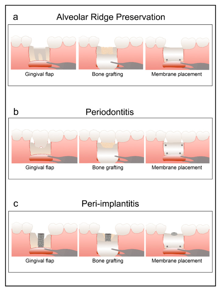
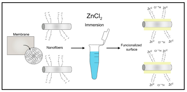
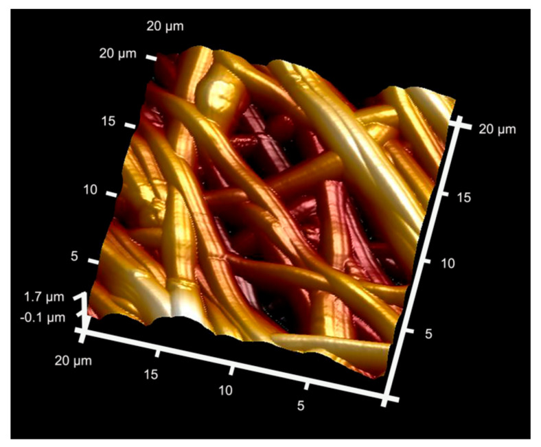
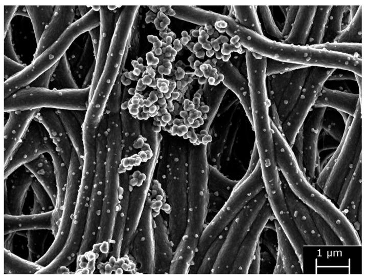
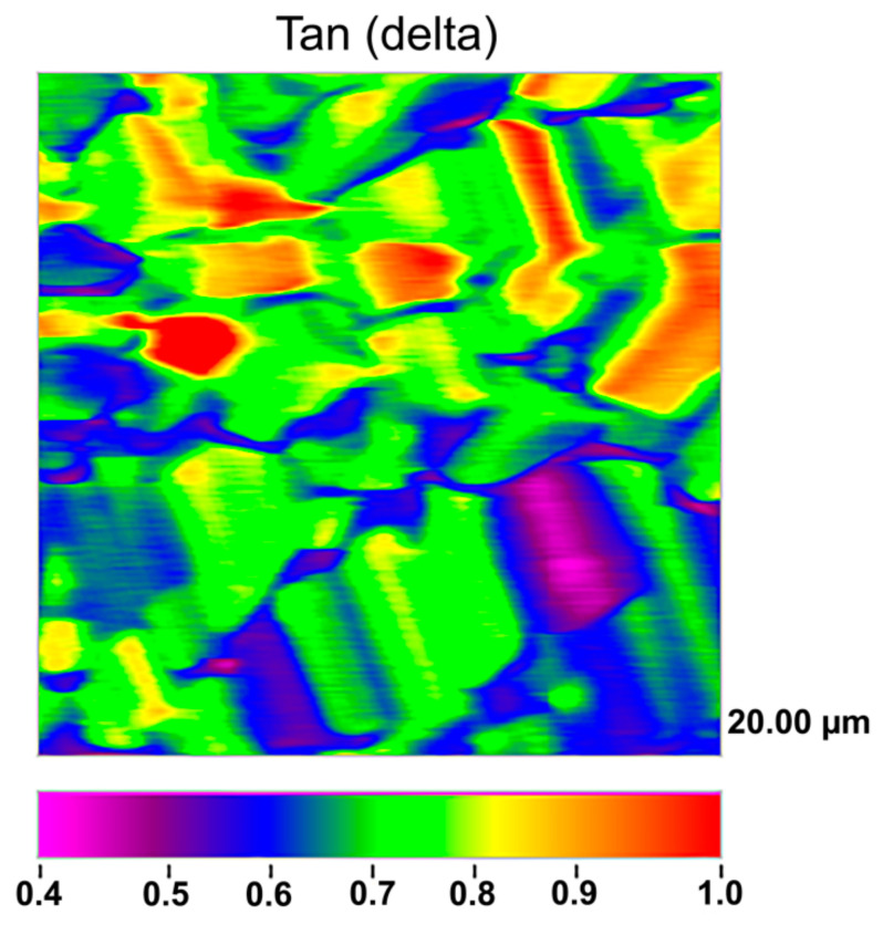
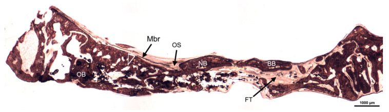
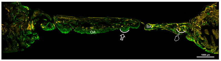
Barrier membranes are employed in guided bone regeneration (GBR) to facilitate bone in-growth. A bioactive and biomimetic Zn-doped membrane with the ability to participate in bone healing and regeneration is necessary. The aim of the present study is to state the effect of doping the membranes for GBR with zinc compounds in the improvement of bone regeneration. A literature search was conducted using electronic databases, such as PubMed, MEDLINE, DIMDI, Embase, Scopus and Web of Science. A narrative exploratory review was undertaken, focusing on the antibacterial effects, physicochemical and biological properties of Zn-loaded membranes. Bioactivity, bone formation and cytotoxicity were analyzed. Microstructure and mechanical properties of these membranes were also determined. Zn-doped membranes have inhibited in vivo and in vitro bacterial colonization. Zn-alloy and Zn-doped membranes attained good biocompatibility and were found to be non-toxic to cells. The Zn-doped matrices showed feasible mechanical properties, such as flexibility, strength, complex modulus and tan delta. Zn incorporation in polymeric membranes provided the highest regenerative efficiency for bone healing in experimental animals, potentiating osteogenesis, angiogenesis, biological activity and a balanced remodeling. Zn-loaded membranes doped with SiO2 nanoparticles have performed as bioactive modulators provoking an M2 macrophage increase and are a potential biomaterial for promoting bone repair. Zn-doped membranes have promoted pro-healing phenotypes.
Keywords: antibacterial; bioactivity; cytotoxicity; guided bone regeneration; mechanical; membranes; microscopy; zinc.
Conflict of interest statement
The authors declare no conflict of interest.
Figures
Similar articles
-
Doxycycline and Zinc Loaded Silica-Nanofibrous Polymers as Biomaterials for Bone Regeneration.Polymers (Basel). 2020 May 25;12(5):1201. doi: 10.3390/polym12051201. Polymers (Basel). 2020. PMID: 32466191 Free PMC article.
-
Polymeric zinc-doped nanoparticles for high performance in restorative dentistry.J Dent. 2021 Apr;107:103616. doi: 10.1016/j.jdent.2021.103616. Epub 2021 Feb 23. J Dent. 2021. PMID: 33636241 Review.
-
A pure zinc membrane with degradability and osteogenesis promotion for guided bone regeneration: In vitro and in vivo studies.Acta Biomater. 2020 Apr 1;106:396-409. doi: 10.1016/j.actbio.2020.02.024. Epub 2020 Feb 22. Acta Biomater. 2020. PMID: 32092431
-
Novel non-resorbable polymeric-nanostructured scaffolds for guided bone regeneration.Clin Oral Investig. 2020 Jun;24(6):2037-2049. doi: 10.1007/s00784-019-03068-8. Epub 2019 Sep 6. Clin Oral Investig. 2020. PMID: 31493213
-
Zinc based biodegradable metals for bone repair and regeneration: Bioactivity and molecular mechanisms.Mater Today Bio. 2023 Dec 28;25:100932. doi: 10.1016/j.mtbio.2023.100932. eCollection 2024 Apr. Mater Today Bio. 2023. PMID: 38298560 Free PMC article. Review.
Cited by
-
Does zinc oxide nanoparticles potentiate the regenerative effect of platelet-rich fibrin in healing of critical bone defect in rabbits?BMC Vet Res. 2022 Apr 2;18(1):130. doi: 10.1186/s12917-022-03231-6. BMC Vet Res. 2022. PMID: 35366880 Free PMC article.
-
Biodegradable Zn-5Dy Alloy with Enhanced Osteo/Angio-Genic Activity and Osteointegration Effect via Regulation of SIRT4-Dependent Mitochondrial Function.Adv Sci (Weinh). 2024 Apr;11(13):e2307812. doi: 10.1002/advs.202307812. Epub 2024 Jan 19. Adv Sci (Weinh). 2024. PMID: 38243646 Free PMC article.
-
Physical cues in biomaterials modulate macrophage polarization for bone regeneration: a review.Front Bioeng Biotechnol. 2025 Jul 23;13:1640560. doi: 10.3389/fbioe.2025.1640560. eCollection 2025. Front Bioeng Biotechnol. 2025. PMID: 40771725 Free PMC article. Review.
-
Histomorphometric Analysis of Differential Regional Bone Regeneration Induced by Distinct Doped Membranes.Polymers (Basel). 2022 May 19;14(10):2078. doi: 10.3390/polym14102078. Polymers (Basel). 2022. PMID: 35631960 Free PMC article.
-
Evaluation of angiogenic, osteogenic, and antimicrobial potential of novel type X collagen-derived GBR membrane: an in vivo preliminary study.Odontology. 2025 Aug 9. doi: 10.1007/s10266-025-01163-9. Online ahead of print. Odontology. 2025. PMID: 40783478
References
-
- Schropp L., Wenzel A., Kostopoulos L., Karring T. Bone Healing and Soft Tissue Contour Changes Following Single-Tooth Extraction: A Clinical and Radiographic 12-Month Prospective Study. Int. J. Periodontics Restor. Dent. 2003;23:313–323. - PubMed
-
- Sheikh Z., Sima C., Glogauer M. Bone Replacement Materials and Techniques Used for Achieving Vertical Alveolar Bone Augmentation. Materials. 2015;8:2953–2993. doi: 10.3390/ma8062953. - DOI
-
- Zafar M.S., Khurshid Z., Almas K. Oral Tissue Engineering Progress and Challenges. Tissue Eng. Regen. Med. 2015;12:387–397. doi: 10.1007/s13770-015-0030-6. - DOI
Publication types
Grants and funding
LinkOut - more resources
Full Text Sources
