

Application of Advanced Nanomaterials for Kidney Failure Treatment and Regeneration
- PMID: 34072461
- PMCID: PMC8198057
- DOI: 10.3390/ma14112939
Application of Advanced Nanomaterials for Kidney Failure Treatment and Regeneration
Abstract
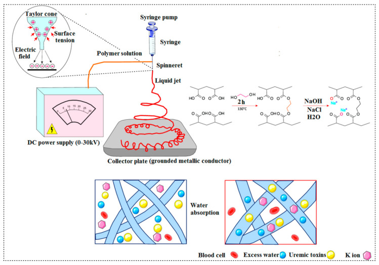
The implementation of nanomedicine not only provides enhanced drug solubility and reduced off-target adverse effects, but also offers novel theranostic approaches in clinical practice. The increasing number of studies on the application of nanomaterials in kidney therapies has provided hope in a more efficient strategy for the treatment of renal diseases. The combination of biotechnology, material science and nanotechnology has rapidly gained momentum in the realm of therapeutic medicine. The establishment of the bedrock of this emerging field has been initiated and an exponential progress is observed which might significantly improve the quality of human life. In this context, several approaches based on nanomaterials have been applied in the treatment and regeneration of renal tissue. The presented review article in detail describes novel strategies for renal failure treatment with the use of various nanomaterials (including carbon nanotubes, nanofibrous membranes), mesenchymal stem cells-derived nanovesicles, and nanomaterial-based adsorbents and membranes that are used in wearable blood purification systems and synthetic kidneys.
Keywords: carbon nanotubes; electrospinning; exosomes; kidney regeneration; nanofibers; nanovesicles; synthetic kidney; therapeutic nanomedicine.
Conflict of interest statement
The authors declare no conflict of interest.
Figures
Similar articles
-
The Clinical Translation of Organic Nanomaterials for Cancer Therapy: A Focus on Polymeric Nanoparticles, Micelles, Liposomes and Exosomes.Curr Med Chem. 2018;25(34):4224-4268. doi: 10.2174/0929867324666170830113755. Curr Med Chem. 2018. PMID: 28875844 Review.
-
Nanomaterials for Nanotheranostics: Tuning Their Properties According to Disease Needs.ACS Nano. 2020 Mar 24;14(3):2585-2627. doi: 10.1021/acsnano.9b08133. Epub 2020 Feb 17. ACS Nano. 2020. PMID: 32031781 Review.
-
Nanomedicine in cancer stem cell therapy: from fringe to forefront.Cell Tissue Res. 2018 Dec;374(3):427-438. doi: 10.1007/s00441-018-2928-5. Epub 2018 Oct 9. Cell Tissue Res. 2018. PMID: 30302547 Review.
-
Material Characterization and Bioanalysis of Hybrid Scaffolds of Carbon Nanomaterial and Polymer Nanofibers.ACS Omega. 2019 Mar 31;4(3):5044-5051. doi: 10.1021/acsomega.9b00197. Epub 2019 Mar 8. ACS Omega. 2019. PMID: 30949614 Free PMC article.
-
Application of nanotubes and nanofibres in nerve repair. A review.Folia Neuropathol. 2010;48(4):231-7. Folia Neuropathol. 2010. PMID: 21225505 Review.
Cited by
-
Patients with Different Stages of Chronic Kidney Disease Undergoing Intravenous Contrast-Enhanced Computed Tomography-The Incidence of Contrast-Associated Acute Kidney Injury.Diagnostics (Basel). 2022 Mar 30;12(4):864. doi: 10.3390/diagnostics12040864. Diagnostics (Basel). 2022. PMID: 35453910 Free PMC article.
-
COUP-TFII in Kidneys, from Embryos to Sick Adults.Diagnostics (Basel). 2022 May 9;12(5):1181. doi: 10.3390/diagnostics12051181. Diagnostics (Basel). 2022. PMID: 35626336 Free PMC article. Review.
-
Incorporating knowledge of disease-defining hub genes and regulatory network into a machine learning-based model for predicting treatment response in lupus nephritis after the first renal flare.J Transl Med. 2023 Feb 3;21(1):76. doi: 10.1186/s12967-023-03931-z. J Transl Med. 2023. PMID: 36737814 Free PMC article.
-
Blood Biomarkers and Metabolomic Profiling for the Early Diagnosis of Vancomycin-Associated Acute Kidney Injury: A Systematic Review and Meta-Analysis of Experimental Studies.J Pers Med. 2022 Aug 28;12(9):1397. doi: 10.3390/jpm12091397. J Pers Med. 2022. PMID: 36143182 Free PMC article. Review.
-
Electric field-directed migration of mesenchymal stem cells enhances their therapeutic potential on cisplatin-induced acute nephrotoxicity in rats.Naunyn Schmiedebergs Arch Pharmacol. 2023 Jun;396(6):1077-1093. doi: 10.1007/s00210-022-02380-7. Epub 2023 Jan 14. Naunyn Schmiedebergs Arch Pharmacol. 2023. PMID: 36640200 Free PMC article.
References
Publication types
Grants and funding
LinkOut - more resources
Full Text Sources
