. 2021 May 29;10(11):2417.
doi: 10.3390/jcm10112417.

60-S Retrogated Compressed Sensing 2D Cine of the Heart: Sharper Borders and Accurate Quantification

Affiliations

60-S Retrogated Compressed Sensing 2D Cine of the Heart: Sharper Borders and Accurate Quantification

Benjamin Longère et al. J Clin Med. .

Abstract

Background and objective: Real-time compressed sensing cine (CSrt) provides reliable quantification for both ventricles but may alter image quality. The aim of this study was to assess image quality and the accuracy of left (LV) and right ventricular (RV) volumes, ejection fraction and mass quantifications based on a retrogated segmented compressed sensing 2D cine sequence (CSrg).

Methods: Thirty patients were enrolled. Each patient underwent the reference retrogated segmented steady-state free precession cine sequence (SSFPref), the real-time CSrt cine and the segmented retrogated prototype CSrg sequence providing the same slices. Functional parameters quantification and image quality rating were performed on SSFPref and CSrg images sets. The edge sharpness, which is an estimate of the edge spread function, was assessed for the three sequences.

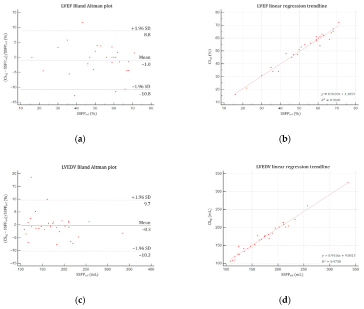
Results: The mean scan time was: SSFPref = 485.4 ± 83.3 (SD) s (95% CI: 454.3-516.5) and CSrg = 58.3 ± 15.1 (SD) s (95% CI: 53.7-64.2) (p < 0.0001). CSrg subjective image quality score (median: 4; range: 2-4) was higher than the one provided by CSrt (median: 3; range: 2-4; p = 0.0008) and not different from SSFPref overall quality score (median: 4; range: 2-4; p = 0.31). CSrg provided similar LV and RV functional parameters to those assessed with SSFPref (p > 0.05). Edge sharpness was significantly better with CSrg (0.083 ± 0.013 (SD) pixel-1; 95% CI: 0.078-0.087) than with CSrt (0.070 ± 0.011 (SD) pixel-1; 95% CI: 0.066-0.074; p = 0.0004) and not different from the reference technique (0.075 ± 0.016 (SD) pixel-1; 95% CI: 0.069-0.081; p = 0.0516).

Conclusions: CSrg cine provides in one minute an accurate quantification of LV and RV functional parameters without compromising subjective and objective image quality.

Keywords: CMR; cardiac; compressed sensing; fast imaging; function; heart; image quality; magnetic resonance; retrogating; retrospective.


Conflict of interest statement

B.L.; C.V.G; A.C.; L.G.; V.S.; J.P.; A.S.; J.H.; D.M.; F.P. have no competing interest. They are employed by an institution engaged in a contractual collaboration with Siemens Healthcare. M.S.; C.F.; S.T. are employees of Siemens Healthcare GmbH.

Figures

Figure 1
Figure 1
Edge sharpness measurement. An orthogonal profile line is drawn across the border between the interventricular myocardium and the left ventricular blood pool (blue line) on a 4-chamber view at end-diastole provided by (a) the reference segmented SSFP sequence, (b) the real-time compressed sensing sequence and (c) the retrogated compressed sensing prototype, providing intensity profiles (blue curves) along the line for (d) reference, (e) real-time and (f) prototype sequences. The edge sharpness was calculated as the inverse of the distance d (in pixels) from the positions corresponding to 20% and 80% (red stars) of the difference between the maximum and minimum signal intensities (black crosses) along the profile line and was expressed in pixel−1. Abbreviations: SSFPref, reference steady-state free precession; CSrt, real-time compressed sensing; CSrg, retrogated compressed sensing; ε, edge sharpness; Ymax, maximum signal intensity; Ymin, minimum signal intensity; d, distance along the intensity (pixels).
Figure 2
Figure 2
Midventricular short-axis cine slice acquired with the three cine sequences in a 37-year-old patient referred for myocarditis suspicion. (a) Reference steady-state free precession cine: Likert scale = 4/4; EuroCMR score = 0/10; ε = 0.082 pixel−1; LVEF = 59%, LVEDV = 135 mL, LVM = 83 g, RVEF = 63%, RVEDV = 159 mL; (b) Real-time compressed sensing cine: Likert scale = 4/3; EuroCMR score = 0/10; ε = 0.056 pixel−1; LVEF = 60%, LVEDV = 133 mL, LVM = 81 g, RVEF = 61%, RVEDV = 153 mL; (c) Retrogated compressed sensing cine: Likert scale = 4/4; EuroCMR score = 0/10; ε = 0.081 pixel−1; LVEF = 58%, LVEDV = 133 mL, LVM = 86 g, RVEF = 62%, RVEDV = 164 mL. Abbreviations: ε, edge sharpness; LVEF, left ventricular ejection fraction; LVEDV, left ventricular end-diastolic volume; LVM, left ventricular myocardial mass; RVEF, right ventricular ejection fraction; RVEDV, right ventricular end-diastolic volume.
Figure 3
Figure 3
Edge sharpness comparison assessed at end-diastole. (a) There was no significant difference regarding ε between CSrg and SSFPref (p = 0.0516) (b) but CSrg significantly improved ε while compared with CSrt (p = 0.0004). Abbreviations: CSrg, retrogated compressed sensing; CSrt, real-time compressed sensing; SSFPref, reference steady-state free precession; ε, edge sharpness.
Figure 4
Figure 4
Bland–Altman plots and linear regression trendlines applied to left ventricular functional parameters quantifications. Left column: Bland–Altman plots for (a) LVEF, (c) LVEDV, (e) LVESV, (g) LVSV and (i) LVM. Solid blue lines are the mean differences between parameters measured with SSFPref and CSrg sequences and dashed red lines are the 95% limits of agreement. Right column: linear regression trend lines for (b) LVEF, (d) LVEDV, (f) LVESV, (h) LVSV and (j) LVM. Abbreviations: SSFPref, reference steady-state free precession; CSrg, retrogated compressed sensing; SD, standard deviation; LVEF, left ventricular ejection fraction; LVEDV, left ventricular end-diastolic volume; LVESV, left ventricular end-systolic volume; LVSV, left ventricular stroke volume; LVM, left ventricular mass.
Figure 4
Figure 4
Bland–Altman plots and linear regression trendlines applied to left ventricular functional parameters quantifications. Left column: Bland–Altman plots for (a) LVEF, (c) LVEDV, (e) LVESV, (g) LVSV and (i) LVM. Solid blue lines are the mean differences between parameters measured with SSFPref and CSrg sequences and dashed red lines are the 95% limits of agreement. Right column: linear regression trend lines for (b) LVEF, (d) LVEDV, (f) LVESV, (h) LVSV and (j) LVM. Abbreviations: SSFPref, reference steady-state free precession; CSrg, retrogated compressed sensing; SD, standard deviation; LVEF, left ventricular ejection fraction; LVEDV, left ventricular end-diastolic volume; LVESV, left ventricular end-systolic volume; LVSV, left ventricular stroke volume; LVM, left ventricular mass.
Figure 5
Figure 5
Bland–Altman plots and linear regression trendlines applied to right ventricular functional parameters quantifications. Left column: Bland-Altman plots for (a) RVEF, (c) RVEDV, (e) RVESV, and (g) RVSV. Solid blue lines are the mean differences between parameters measured with SSFPref and CSrg sequences and dashed red lines are the 95% limits of agreement. Right column: linear regression trend lines for (b) RVEF, (d) RVEDV, (f) RVESV, and (h) RVSV. Abbreviations: SSFPref, reference steady-state free precession; CSrg, retrogated compressed sensing; SD, standard deviation; RVEF, right ventricular ejection fraction; RVEDV, right ventricular end-diastolic volume; RVESV, right ventricular end-systolic volume; RVSV, right ventricular stroke volume.

References

    1. Pennell D.J., Sechtem U.P., Higgins C.B., Manning W.J., Pohost G.M., Rademakers F.E., van Rossum A.C., Shaw L.J., Yucel E.K., Society for Cardiovascular Magnetic Resonance et al. Clinical indications for cardiovascular magnetic resonance (CMR): Consensus panel report. Eur. Heart J. 2004;25:1940–1965. doi: 10.1016/j.ehj.2004.06.040. - DOI - PubMed
    1. Maceira A.M., Prasad S.K., Khan M., Pennell D.J. Normalized left ventricular systolic and diastolic function by steady state free precession cardiovascular magnetic resonance. J. Cardiovasc. Magn. Reson. 2006;8:417–426. doi: 10.1080/10976640600572889. - DOI - PubMed
    1. Maceira A.M., Prasad S.K., Khan M., Pennell D.J. Reference right ventricular systolic and diastolic function normalized to age, gender and body surface area from steady-state free precession cardiovascular magnetic resonance. Eur. Heart J. 2006;27:2879–2888. doi: 10.1093/eurheartj/ehl336. - DOI - PubMed
    1. Curtis J.P., Sokol S.I., Wang Y., Rathore S.S., Ko D.T., Jadbabaie F., Portnay E.L., Marshalko S.J., Radford M.J., Krumholz H.M. The association of left ventricular ejection fraction, mortality, and cause of death in stable outpatients with heart failure. J. Am. Coll. Cardiol. 2003;42:736–742. doi: 10.1016/S0735-1097(03)00789-7. - DOI - PubMed
    1. Karamitsos T.D., Francis J.M., Myerson S., Selvanayagam J.B., Neubauer S. The role of cardiovascular magnetic resonance imaging in heart failure. J. Am. Coll. Cardiol. 2009;54:1407–1424. doi: 10.1016/j.jacc.2009.04.094. - DOI - PubMed

LinkOut - more resources