. 2021 May 29;22(11):5833.
doi: 10.3390/ijms22115833.

Role Played by Receptors for Advanced Glycosylation End Products in Corneal Endothelial Cells after HSV-1 Infection

Affiliations

Role Played by Receptors for Advanced Glycosylation End Products in Corneal Endothelial Cells after HSV-1 Infection

Dai Miyazaki et al. Int J Mol Sci. .

Abstract

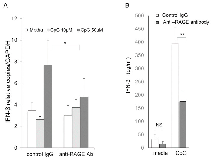
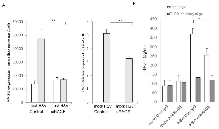
Senescence, sterile inflammation, and infection cause dysfunction of corneal endothelial cells, leading to visual morbidity that may require corneal transplantation. With increasing age, the extracellular matrix is modified by non-enzymatic glycation forming advanced glycation end products (AGEs). The modifications are primarily sensed by the receptors for the AGEs (RAGE) and are manifested as a type I interferon response. Interestingly, in our study, human corneal endothelial cells (HCEn) cells did not respond to the typical RAGE ligands, including the AGEs, high mobility group box 1 (HMGB1), and serum amyloid-A (SAA). Instead, HCEn cells responded exclusively to the CpG DNA, which is possessed by typical corneal pathogen, herpes simplex virus-1 (HSV-1). Upon HSV-1 infection, the surface expression of RAGE was increased, and endocytosed HSV-1 was associated with RAGE and CpG DNA receptor, TLR9. RAGE DNA transfection markedly increased interferon-β secretion by CpG DNA or HSV-1 infection. HSV-1 infection-induced interferon-β secretion was abolished by TLR9 inhibition and partially by RAGE inhibition. Global transcriptional response analysis confirmed that RAGE and TLR9 were both significantly involved in type I interferon responses. We conclude that RAGE is a sensor of HSV-1 infection and provokes a type I interferon response.

Keywords: advanced glycation end products; corneal endothelial cell; herpes simplex virus; receptor for advanced glycosylation end products; toll-like receptor 9.


Conflict of interest statement

The authors declare no competing financial interest.

Figures

Figure 1
Figure 1
Responsiveness of human corneal endothelial (HCEn) cells to the receptors of advanced glycation end products (RAGE) ligands assessed by the level of expression of interferon-β. Exposure to CpG DNA significantly induces interferon-β expression after 12 h (left). Advanced glycation end products (AGE), HMGB1, and serum amyloid A (SAA) (right) do not significantly induce interferon-β. DMEM with 10% FBS was used for the assay media. Left and right panels were obtained from different batches of cells of single origin. For * p < 0.00001; n = 6. Ns: not significant.
Figure 2
Figure 2
Induction of RAGE expression after HSV-1 infection. HCEn cells were infected with HSV-1 at multiplicity of infection (MOI) of 1 and evaluated for the expression of RAGE. (A) The induction of the mRNA of RAGE determined by real-time RT-PCR is significantly increased and peaked at 24 h post infection. *: p < 0.00001. n = 4. (B) Cell surface expression of RAGE after HSV-1 infection was assessed by FACS analysis. RAGE expression is increased after infection and peaked at 24 h. Mean signal intensity of RAGE was 26 ± 1 (mock), 55 ± 3 (6 h, p = 0.001), 117 ± 6 (12 h, p = 0.000), and 149 ± 7 (24 h, p = 0.000). n = 374/group.
Figure 3
Figure 3
Cellular localization of TLR9, RAGE, and HSV-1. HCEn cells were infected with GFP-HSV (at MOI 50, arrow (green)) and stained for cell surface RAGE (arrow (red): PE-labeled) and TLR9 (Alexa647-labeled, arrow (yellow)) without permeabilization. HSV-1 is colocalized with cell surface RAGE, which partly overlapped with the accumulated TLR9 expression (upper panel). Lower panel: GFP-HSV (arrow (green)) was associated with TLR9 (arrow (yellow)) near cell surface. TLR9 expression was transitioned to endoplasmic reticulum (ER, blue) surrounding nucleus (lower panel). Nuclear transition of GFP-HSV is also observed at 3 h PI. Bar indicates 10 µm.
Figure 4
Figure 4
Association of TLR9, RAGE, and HSV-1. (A) Association of HSV-1 and RAGE by a GFP-pull-down assay. HCEn cells were infected with HSV-1 at MOI of 50, and the cell lysates as input were pulled down by anti-GFP antibody. The proteins associated with GFP-HSV-1 were detected by SDS PAGE. SDS PAGE, gel stained by Coomassie Blue, is shown as the input, pull down, and flow through fraction (upper panel). To observe RAGE and TLR9, Western blot for each fraction was conducted for RAGE and TLR9 (lower panel). (B) Tubulin and GFP in each fraction are shown by Western blot.
Figure 5
Figure 5
Network analysis of RAGE (AGER) and HSV-1 infection-induced transcriptional responses of HCEn cells. Functional analysis was conducted to determine the associations of the HSV-1 infection-induced genes with biological functions. A set of 430 infection-induced genes (fold induction > 4) were analyzed for network generation of biological functions. The 3 highest significant networks (p < 1 × 10⁻³²) are summarized as merged networks. RAGE (AGER) and TLR9 were significantly associated with type I interferon responses.
Figure 6
Figure 6
Induction of interferon-β by HCEn cells overexpressing RAGE. HCEn cells were transfected with RAGE-expressing plasmid or control plasmid. Transfection of RAGE c-DNA results in 35kDa, 43kDa, and 70kDa bands by Western blot, reflecting post translational processing [20]. Overexpressed RAGE was shown for 70 kDa band (A). RAGE-transfected HCEn cells were assessed for interferon-β secretion 24 h after CpG exposure or HSV-1 infection. Forced induction of RAGE increased the expression of interferon-β by CpG (50 μM) (B), and HSV infection at multiplicity of infection (MOI) 1 (C). *: p = 0.01, **: p = 0.001, ***: p <0.001; n = 6.
Figure 7
Figure 7
Role of RAGE in interferon-β production by CpG oligonucleotide stimulation of corneal endothelial cells. (A) HCEn cells were stimulated by CpG oligonucleotides and were assessed for interferon-β mRNA induction at 12 h by real-time RT-PCR. HCEn cells stimulated by CpG oligonucleotides significantly induced interferon-β. Inhibition by anti-RAGE antibody significantly reduced the induction of interferon-β by CpG oligonucleotide. (B) HCEn cells were stimulated by CpG and were assessed for interferon-β production at 24 h by ELISA. The interferon-β production by CpG oligonucleotides (50 μM) was significantly reduced by anti-RAGE antibody. n = 6, *: p = 0.01, **: p < 0.001. Ns: not significant.
Figure 8
Figure 8
Role of RAGE and TLR9 in the interferon-β production by HSV-1 infection of corneal endothelial cells. (A) HCEn cells were transfected by siRNA of RAGE or control siRNA, and they were assessed for cell surface expression of RAGE with or without HSV-1 infection (multiplicity of infection (MOI) 1. Plated cells were stained for the surface expression of RAGE, and 1300 cells in high powered field per group were assessed for total fluorescence intensity. Si RNA transfection abolished HSV-1-induced-RAGE expression. HCEn cells were infected by HSV-1 infection at MOI 1 and were assessed for the expression of the mRNA of interferon-β at 12 h by real-time RT-PCR. The HSV-1 infection induced-interferon-β mRNA were significantly impaired by the RAGE blockade using siRAGE. (B) HCEn cells were infected by HSV-1 and were assessed for interferon-β production at 24 h by ELISA. HCEn cells stimulated with HSV-1 infection at MOI 1 induced interferon-β which was significantly reduced by anti-RAGE antibody and abolished by TLR9 inhibitory oligonucleotide (2 µM). *: p < 0.01; **: p <0.001; n = 6.

Similar articles

Cited by

References

    1. Flaxman S.R., Bourne R.R.A., Resnikoff S., Ackland P., Braithwaite T., Cicinelli M.V., Das A., Jonas J.B., Keeffe J., Kempen J.H., et al. Global causes of blindness and distance vision impairment 1990–2020: A systematic review and meta-analysis. Lancet Glob. Health. 2017;5:e1221–e1234. doi: 10.1016/S2214-109X(17)30393-5. - DOI - PubMed
    1. Ung L., Bispo P.J.M., Shanbhag S.S., Gilmore M.S., Chodosh J. The persistent dilemma of microbial keratitis: Global burden, diagnosis, and antimicrobial resistance. Surv. Ophthalmol. 2019;64:255–271. doi: 10.1016/j.survophthal.2018.12.003. - DOI - PMC - PubMed
    1. Garg P., Krishna P.V., Stratis A.K., Gopinathan U. The value of corneal transplantation in reducing blindness. Eye. 2005;19:1106–1114. doi: 10.1038/sj.eye.6701968. - DOI - PubMed
    1. Santos C.Y., Snyder P.J., Wu W.C., Zhang M., Echeverria A., Alber J. Pathophysiologic relationship between Alzheimer’s disease, cerebrovascular disease, and cardiovascular risk: A review and synthesis. Alzheimers Dement. 2017;7:69–87. doi: 10.1016/j.dadm.2017.01.005. - DOI - PMC - PubMed
    1. Howes K.A., Liu Y., Dunaief J.L., Milam A., Frederick J.M., Marks A., Baehr W. Receptor for advanced glycation end products and age-related macular degeneration. Investig. Ophthalmol. Vis. Sci. 2004;45:3713–3720. doi: 10.1167/iovs.04-0404. - DOI - PubMed

MeSH terms

Substances

LinkOut - more resources